
Manuscript accepted on :
Published online on: 18-12-2015
Plagiarism Check: Yes
Vadod Norouzi1, Ali Mohammadian1 and Behzad Eskandaroghli2*
1Department of Anesthesiology, Ardabil University of Medical Science, Ardabil, Iran.
2Department of Neurosurgery, Ardabil University of Medical Science, Ardabil, Iran.
DOI : https://dx.doi.org/10.13005/bpj/412
Abstract
The present study aimed to determine the relationship between initial and 24 h base deficit after the patient rehabilitation in critically ill inpatients in the surgical ICU to determine the prognosis of patients. 96 patients hospitalized in the surgical ICU during 6 months were enrolled regardless of the reason for admission. The statistical correlation between initial and 24 h base deficit and the patients’ outcome were examined. The raw data were analyzed using descriptive and analytical statistical methods by SPSS. Various statistical tests such as T-test, Chi-Square and Roc curve were used. The results with P-value less than 0.05 were statistically considered valid. The initial and 24 h base deficit of patients with death outcome was significantly higher than those with discharge outcome (P<0.001). The initial arterial pH had no statistically significant correlation with patients’ outcome (p=0.46). In contrast, the 24 h arterial pH was associated with outcome (p= 0.066). There was a poor correlation between arterial blood pressure at admission and patients’ outcome (p=0.06). The values of initial and 24h base deficit with the most predictor ability of patients’ outcome were -11.35 and -4.6. Accordingly, the deficit of initial base up to -11.35 has been associated with mortality rate of 19.7%. Values less than -11.35 has been associated with mortality rate of 65.4% (p=0.001). Furthermore, the 24 h base deficit up to -4.6 has been associated with mortality rate of 15.2%. Values less than -4.6 has been associated with mortality rate of 61.5% (p<0.001). The increased levels of initial and 24 h base deficit have a strong correlation with the outcome of critically ill patients. Therefore, it can be used in determining the prognosis of critically ill patients.
Keywords
Base Deficit; Outcome; the Intensive Care
Download this article as:| Copy the following to cite this article: Norouzi V, Mohammadian A, Eskandaroghli B. The Initial and 24 h (After the Patient Rehabilitation) Deficit of Arterial Blood Gases as Predictors of Patients Outcome. Biomed Pharmacol J 2013;6(2) |
| Copy the following to cite this URL: Norouzi V, Mohammadian A, Eskandaroghli B. The Initial and 24 h (After the Patient Rehabilitation) Deficit of Arterial Blood Gases as Predictors of Patients Outcome. Biomed Pharmacol J 2013;6(2). Available from: http://biomedpharmajournal.org/?p=2714 |
Introduction
Quickly diagnosis and improvement of physiological disorders is a key component in the management of critically ill patients [1]. Damages caused by accidents as well as surgical emergencies both can cause serious physiological and metabolic disorders. If not treated properly, they can cause dysfunction of vital organs or complete loss of function and ultimately death [2].
In the medical care of critically ill patients, early rehabilitation of the patient is critical for going out of shock and more quickly tissue hypoxia improvement [3]. The response to initial rehabilitation at the bedside of critically ill patients is usually evaluated based on the clinical findings such as arterial blood pressure, heart rate and urinary flow. These clinical evidence are poor criteria in terms of studying the return of tissue hypoxia process [4]. In other words, the patient’s vital signs are maintained in normal range with the intervention of several physiological mechanisms. But the shock and tissue hypoxia is thriving and it seems that it will be the main factor determining the prognosis of patients in terms of complications and mortality.
Substantially disrupt of the physiological balance and reduce of perfusion to organs and thereby metabolic acidosis is the main concern in these patients. In fact, the metabolic acidosis is the key factor in the formation of the terrible triad of acidosis, hypothermia and coagulopathy [5]. Thus, the measurement of local markers or systemic metabolic acidosis has been widely used in the assessment of severity of injury or illness, treatment adequacy and prognosis [3].
In this regard, researchers have looked for a criterion and tool to precisely and quantitatively evaluate the tissue hypoxia and tried to improve it. The recent studies in this field proposed the serum lactate and base deficit levels as an indicator of anaerobic metabolism and tissue hypoxia [6]. In fact, these two criteria can be quickly calculated by measuring and analyzing the arterial blood gas at the bedside, accordingly the patient recovery will be followed .[3] Arterial base deficit is a quantitative value calculated based on arterial pressure of CO2, HCO3 and pH. It represents the amount of additional equivalent base which must be added to 1 liter of blood to normalize the pH of blood [7].
Smith et al. (2001) conducted a descriptive-prospective study on 148 inpatients in the general ICU. They considered base and serum lactate deficit as prognosis indicators in patients hospitalized in the ICU. Both criteria of base and serum lactate deficit have been invaluable in determining the prognosis of patients. The combination of these two criteria had the sensitivity and specificity of 80% and 58% for mortality, respectively [8].
Farah et al (2003) conducted a retrospective study on 137 inpatients in the surgical ICU to investigate the relationship between base deficit and serum lactate level at admission and 24 h later and the disease outcome. They suggested that base deficit is as poor indicator to determine the prognosis in all patients. In contrast, the initial serum lactate level has been more valuable to determine prognosis. They also studied the relationship between the time required to reach the serum lactate to normal level and disease outcome. So that in the case where the level of serum lactate reached to normal level within 24 h after admission, the mortality rate was 10%, while in the case where the serum lactate reached to normal level within more than 48 h later, mortality rate was 67% [3].
Martin et al. (2006) conducted a study entitled “the discordance in base and lactate deficit in determining prognosis in surgical ICU patients”. In this study, base and lactate level deficit on admission have been introduced as a marker of metabolic acidosis and the prognosis determination. But the increase in base deficit alone without increased serum lactate level had a little predictive value. In contrast, increased level of serum lactate has been associated with higher rates of mortality and complications even without an increase in base deficit [1].
FitzSullivan et al. (2005) conducted a study entitled “serum bicarbonate as an alternative for arterial base deficit in trauma patients”. They examined 3102 patients admitted to the surgical ICU during 1996-2004 who had required experimental values. The relationship between serum level of HCO3– and base deficit was analyzed by statistical methods. The serum HCO3– level had a direct correlation with base deficit. Moreover, the value of serum bicarbonate in determining patients’ prognosis of arterial base deficit was determined. According to the results of this study, to determine the prognosis, measurement of the serum HCO3– can easily be replaced with base deficit [9].
Schmelzer et al. (2008) conducted a prospective study on 100 inpatients in the surgical ICU entitled “a comparison of central venous and arterial base deficit as a predictor of survival in acute trauma”. Demographic data of patients, injury severity score (ISS), venous and arterial base deficit, pH, Pco2, Po2, HCO3 and outcome of patients were measured. Of these patients, 76 people have been treated and 26 patients died. Venous not arterial base deficit was introduced as a predictor of patients’ prognosis [10].
The present study aims to examine the relationship between initial base deficit and 24 h after the patient rehabilitation and the prognosis in traumatic and non-traumatic patients hospitalized in surgical ICU.
Materials and Methods
This study was conducted on patients hospitalized in surgical ICU of Fatemi Hospital regardless of the cause of hospitalization or initial diagnosis. Inclusion criteria were the presence of ABG values at admission and 24 h later in the patient’s records and at least 24 h stay in the ICU.
By referring admission and discharge registering book of patients in ICU, 96 inpatients were enrolled taking into account the inclusion criteria from April to September 2009. Patients were hospitalized with a physician in consultation with the anesthesiologist. Among these patients, the initial ABG results were fully recorded in the records of 87 patients. These cases were statistically analyzed.
The requited information was extracted by designing a questionnaire containing demographic characteristics of the patients, vital signs at admission, initial tests, initial BD value and its value 24h after admission, duration of hospitalization and patient outcomes referring to the records of inpatients. Patients’ vital signs were obtained according to the report of doctor or nurses. The expected outcome of patients including discharge, mortality and complications during the hospitalization were extracted based on the contents of the records.
The patients were examined in terms of complications during hospitalization including acute renal failure, oliguria, the need for dialysis, pulmonary complications such as the need for artificial ventilation, pulmonary infections, adverse cardiovascular outcomes such as the need for the use of single-vessel contractors, acute myocardial infarction, heart dysrhythmia and septic complications including wound infection, sepsis and urinary tract infection. The obtained data were entered into the questionnaire. Initial resuscitation of patients in the ED was performed according to the emergency physician. The treatment was followed by a surgeon doctor or an anesthesiologist during hospitalization in ICU.
The patients were evaluated for the presence of comorbidities. None of the patients had underlying disease affecting the metabolism and ABG results. ABG parameters, in addition to the BD, including HCO3, CO2, and pH were also extracted. The date of ABG was obtained according to the nursing report or the date stated in the analysis. In this study, arterial ABG results were analyzed. The raw data were analyzed using descriptive statistical methods by SPSS. Different statistical tests such as T-test, Chi-Square and Roc curve were employed. The statistical validity of results was considered with the value P <0.005.
Table 1: The significance of the relationship of various variables with patients’ outcome
| Outcome Variable | Number | Average | P value |
| Death age
Discharge |
30
62 |
47.53
39.59 |
0.07 |
| SBP Death
Discharge |
29
58 |
91.9
100.6 |
0.06 |
| Death hospitalization duration
Discharge |
27
60 |
4.66
9.43 |
0.001 |
| initial pH Death
Discharge |
29
58 |
7.37
7.28 |
0.46 |
| Initial BD Death
Discharge |
29
58 |
-12.7
-4.1 |
0.001> |
| 24h BD Death
Discharge |
21
38 |
-7.8
-1.13 |
0.001> |
| 24h pH Death
Discharge |
21
38 |
7.31
7.36 |
0.066 |
| Hb Death
Discharge |
25
53 |
11.66
2 |
0.58 |
| BUN Death
Discharge |
23
43 |
50.09
23.4 |
0.019 |
Results
With the quick identification of critically ill patients who require special cares and employing appropriate remedial measures, the mortality of patients can be reduced and the patient’ outcome will be improved. So far, various physiological variables have been introduced for this purpose. This is a quantitative study to clarify the scope of application of one of the above criteria. Overall, 96 patients were enrolled. 76% of patients (n = 73) were male, and 24% (n = 23) were female. Mean age of patients was 42.8 years. 74 patients were traumatic and 22 were non-traumatic cases (Fig. 1).
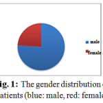 |
Figure 1: The gender distribution of patients (blue: male, red: female) |
Of the 96 patients, 30 (31%) have died including 25.7% of traumatic and 50% of non-traumatic patients. The average initial base deficit in all patients was -7.29. The initial base deficit in traumatic and non-traumatic patients was -6.02 and -12.14, respectively. The average stay in the ICU was 7.8 days. The average stay of traumatic and non-traumatic patients was 8 and 6.50 days, respectively. Table 1 shows the significance of the relationship of the various variables with patients’ outcome. The value of P <0.05 was considered statistically significant.
Based on Table 1, 24h pH, BUN levels, duration of stay and especially the initial and 24h base deficit were statistically significant in determining the patients’ outcome. According to ROC curve, the initial and 24h base deficit of -11.35 and -4.6 respectively were determined as the best prognosis predictors. Based on the ROC curve, mortality rate of patients with initial base deficient less than or equal to -11.35 was 65.6%. In contrast, the initial base deficit greater than -11.35 has been associated with mortality rate of 19.6%. Accordingly, 24 h base deficit less and greater than -4.6 has been associated with mortality rates of 61.5% and 15.2%, respectively. Due to the importance of 24-h base deficit in evaluating the response of patients to the rehabilitation, the patients’ outcome was studied based on 24 h base deficit considering the initial base deficit. This is indicated in Table 2. (Table 2)
Table 2: The outcome of patients based on 24 h base deficit considering initial base deficit
| Initial base deficit 24 h base deficit | Number | percentage | ||
| <-11.35
|
6 4. <-
4.6-< |
Death
Discharge Death Discharge |
11
4 2 4 |
73.3
26.7 33.3 66.7 |
| <-11.35
|
<-4.6
>-4.6 |
Death Discharge
Death Discharge |
5
6 3 24 |
45.5
54.5 11.1 88.9 |
| Total | 87 | 100 | ||
Discussion and Conclusion
The base deficit is an experimental criterion which is easily and quickly measured by blood gas analyzers. In this study, the relationship between initial arterial pH and the outcome of patients was not statistically significant (p= =0.46). In contrast, the initial base deficit was significantly correlated with outcome of patients (p<0/001). Previous studies introduced base deficit as an excellent criterion compared with blood pH to assess cellular hypoxia and shock as well as determining prognosis [8].
In the present study, the normal range of base deficit was determined between 2 to +2 [3]. Accordingly, nearly 70% of patients had abnormal base deficit. The overall mortality rate of patients (31.3%) and the mean stay in the ICU (about 8 days) indicate the critically ill patients.
Davis et al. classified the base deficit in the three categories of moderate (-3 to -5), medium (-6 to -14) and severe (less than -15). They showed that the more negative rates of base deficit, requiring more fluid for initial resuscitation of patients, and increased complications during hospitalization [11].
In this study, based on the ROC curve, the base deficit of -11.35 on admission and -4.6 after 24 h of admission have been introduced as the strongest predictors of patients’ outcome. Accordingly, the initial base deficit less and greater than -11.35 was associated with mortality rate of 19.7% and 65.4%, respectively (p=0.001). Moreover, the 24 h base deficit less and greater than -4.6 was associated with mortality rate of 15.2% and 61.5%, respectively (p <0/001).
According to ROC curve, Smith et al. introduced the initial base deficit less than -4 as the strongest factor associated with mortality. In their study, the more negative initial base deficit of -4 was associated with mortality rate of 57.1%. In contrast, patients with a base deficit greater than or equal to -4 had a mortality rate of 17.6% (p<0.0001) [3].
It seems that the different values of base deficit based on ROC curve in this study with previous studies is justified with the rate of critically ill patients. Ill patients will have a more negative base deficit values. However, it must be noted that the tissue hypoperfusion is not the just cause of metabolic acidosis and base deficit in critically ill patients, but vasopressor administration, normal saline, DKA and increasing cellular metabolism in the context of sepsis or burns are considered as the main causes of metabolic acidosis [8].
In this study, the initial vital signs of patients had a poor correlation with prognosis (p=0.06). This issue has been expressed in previous studies [12]. In some studies, in addition to base deficit, measurement of serum lactate levels was used to determine the prognosis. Farah et al. showed in a study that base deficit is useful in determining the prognosis only in conjunction with hyperlactatemia [3].
Farah et al found no significant correlation between initial base deficit and determination of disease outcome. In this study, only 24 h base deficit was associated with prognosis (p=0.02). In this study, 77% of patients were traumatic. The mortality rate in traumatic and non-traumatic patients was 25.7% and 50%, respectively. The mean duration of hospitalization of traumatic and non-traumatic patients was 8 and 6.5 days, respectively. Initial base deficit in traumatic patients had a significant correlation with disease outcome (p<0.05). The mean base deficit of traumatic and non-traumatic patients was -6.02 and -12.1, respectively. It seems that more negative initial base deficit in non-traumatic patients compared with traumatic patients can justify more mortality rate in this group of patients.
The important point is the improvement of the prognosis of patients who had normal 24 h base deficit after admission. In fact, the effective rehabilitation of this group of patients improved tissue perfusion and resolution of cellular hypoxia and thus reduce the production of acid. Patients with initial base deficit less than -11.35, with 24 h base deficit less and greater than -4.6, had mortality rate of 73.3% and 33.3%, respectively.
In some studies, both arterial and venous base deficit have been compared. Schmelzer et al. (2008) showed that only venous not arterial base deficit is associated with the outcome of patients [10]. In the present study, only the values of arterial base deficit were examined. The results showed that arterial base deficit had a significant correlation with the outcome of patients. As a result, increased levels of initial and 24 h base deficit has a strong correlation with the outcome of critically ill patients. Therefore, it can be used in determining the prognosis of critically ill patients. It seems that the measurement and analysis of base deficit should be considered in the triage and treatment approach of critically ill patients.
References
- Martin, M.J., et al., Discordance between lactate and base deficit in the surgical intensive care unit: which one do you trust? The American journal of surgery, 2006. 191(5): p. 625-630.
- Oestern, H.-J., et al., Cardiorespiratory and metabolic patterns in multiple trauma patients. Resuscitation, 1979. 7(3): p. 169-183.
- Husain, F.A., et al., Serum lactate and base deficit as predictors of mortality and morbidity. The American journal of surgery, 2003. 185(5): p. 485-491.
- Scalea, T.M., et al., Resuscitation of multiple trauma and head injury: role of crystalloid fluids and inotropes. Critical care medicine, 1994. 22(10): p. 1610-1615.
- Cosgriff, N., et al., Predicting life-threatening coagulopathy in the massively transfused trauma patient: hypothermia and acidoses revisited. The Journal of Trauma and Acute Care Surgery, 1997. 42(5): p. 857-862.
- Davis, J.W., The relationship of base deficit to lactate in porcine hemorrhagic shock and resuscitation. The Journal of Trauma and Acute Care Surgery, 1994. 36(2): p. 168-172.
- Maciel, A.T. and M. Park, Differences in acid-base behavior between intensive care unit survivors and nonsurvivors using both a physicochemical and a standard base excess approach: a prospective, observational study. Journal of critical care, 2009. 24(4): p. 477-483.
- Smith, I., et al., Base excess and lactate as prognostic indicators for patients admitted to intensive care. Intensive care medicine, 2001. 27(1): p. 74-83.
- FitzSullivan, E., et al., Serum bicarbonate may replace the arterial base deficit in the trauma intensive care unit. The American journal of surgery, 2005. 190(6): p. 961-967.
- Schmelzer, T.M., et al., A comparison of central venous and arterial base deficit as a predictor of survival in acute trauma. The American journal of emergency medicine, 2008. 26(2): p. 119-123.
- Davis, J.W., et al., Admission base deficit predicts transfusion requirements and risk of complications. The Journal of Trauma and Acute Care Surgery, 1996. 41(5): p. 769-774.
- Luna, G.K., A.C. Eddy, and M. Copass, The sensitivity of vital signs in identifying major thoracoabdominal hemorrhage. The American journal of surgery, 1989. 157(5): p. 512-515.

