
Manuscript accepted on :June 04, 2010
Published online on: 20-11-2015
Plagiarism Check: Yes
J. Malathi* and P. Somashekhar
Department of Microbiology, Shivani college of Pharmacy, Warangal - 506 001, India.
Abstract
The cancer is major disease which is convulsing all the global, so many of hazardous effects carried by this disease. Normally the cancer development occurs due to unregulated body metabolisms, Mutations, Uncontrolled cell growth, some of the proteins also involved in development of cancer disease. The oncogenes are plays vital role in expansion of cancer, normally most cancers are somatic, not germ line mutational events, although some cancers have heritable form, cancer occurrence is sporadic in most cases. Over time, several somatic mutations accumulate with in the same somatic cell lineage, altering or knocking out the functions of several genes, until finally a cancerous cell expand in to a clone of cancerous cells. Addition to this so many of mechanisms converting normal cell in to cancer cell sequential biochemical events depend on the successful occurrence of prior events, in the cell cycle, fail-safe mechanisms prevent the cell cycle from progressing until prior events have been successfully completed, survival factors block advancement of the death pathways. The magnification of cancer is initiated by the various components of the intercellular signaling systems, modified by protein phosphorylation, allosteric interactions between proteins and small molecules, and interaction between protein subunits. The variation also occurs in the amount of albumins and globulins from a normal cell to cancerous cell, where as all the proteins were involved in improvement of disease. The present work is conducted to know the different amounts of serum albumins and globulins present in the cancer patients, for this we compiled 15 positive cancer samples from Warangal district hospital, and subjected to Organic solution precipitation and salt precipitation. Subsequently these precipitated protein samples were analyzed by SDS Polyacrylamide gel electrophoresis, and different bands of albumin and globulin seen with different concentrations in a densitometer.
Keywords
Plasma proteins; a; b; g Globulins; Precipitation; Coomassie blue staining; SDS-PAGE
Download this article as:| Copy the following to cite this article: Malathi J, Somashekhar P. Separation of Cancer Proteins using Polyacrylamide Gel Electrophoresis. Biomed Pharmacol J 2010;3(1) |
| Copy the following to cite this URL: Malathi J, Somashekhar P. Separation of Cancer Proteins using Polyacrylamide Gel Electrophoresis. Biomed Pharmacol J 2010;3(1). Available from: http://biomedpharmajournal.org/?p=1116 |
Introduction
The cancer is one of the major causes of death in the developed nations –at least one in five of the population of Europe and North America can expect to die of cancer. Figures for the last 100 years or so give the impression that the disease is increasing in these countries, but cancer is largely a disease of the later age groups, and with the advances of public health and medical science many more people live to the age where they are able to get cancer.
The terms of cancer, malignant neoplasm and malignant neoplasm and tumors are synonymous; they are distinguished from benign tumours’ by the properties of dedifferentiation, invasiveness and ability to metastasise. The appearance of these characteristics reflects altered patterns of gene expression in the cancer cells, resulting from genetic mutations (1).
There have been numerous advances in the understanding of the pathogenesis of cancer, cancer cells manifest, to varying degrees, four characteristics that distinguish them from normal cells: uncontrolled proliferation, dedifferentiation and loss of function, invasiveness and metastasis (2).
The genesis of a cancer cell
A normal cell turns into a cancer cell because of one or more mutations in its DNA, which can be inherited or acquired (3). The development of cancer is a complex multistage process, involving not only more than one genetic change but usually also other, epigenetic factors (hormonal action, co-carcinogen and tumour promoter effects etc.)(4) that are not in themselves cancer producing but which increase the likelihood that the genetic mutations will result in cancer, there are two main categories of genetic change that lead to cancer: the activation of proto oncogenes to oncogenes and the inactivation of tumour suppressor genes . These changes are result of point mutations, gene amplification or chromosomal translocation, often due to the action of certain viruses or chemical carcinogens (5).
The proliferation of cancer cells is not controlled by the process that normally regulates cell division and tissue growth. It is this, rather than their rate of proliferation, that distinguishes them from normal cells. Some normal cells (such as neurons) have little or no capacity to divide and proliferate, but others, for example in the bone marrow and the epithelium of the gastrointestinal tract, have the property of continuous rapid division (. Some cancer cells multiply slowly (e.g. those in plasma cell tumours) and some fast (e.g. the cells of Burkitt’s lymphoma). It is, there fore, not generally true that cancer cells proliferate faster than normal cells. The significant point about cancer cells us not that they proliferate faster than normal cells but that their proliferation is not subject to normal regulatory process. Many growth factors and inhibitors result in uncontrolled proliferation. The growth factor pathways-the cytosolic and nuclear transducers, the cell cycle transducers-e.g. cyclins, cyclin-dependent kinases (cdks) or the cdk inhibitors. Potentially all the genes coding for apoptotic mechanism, telomerase expression and local blood vessels could be regarded as oncogenes or tumour suppressor genes though not all are equally prone to prone to malignant transformation, and it is obvious that malignant transformation of several components is need for the development of cancer (6).
Apoptosis and the Genesis of Cancer cell
Apoptosis is programmed cell death and anti apoptotic genetic lesions are necessary for cancer to develop. In fact development of resistance to apoptosis is a hallmark of cancer. Decreased apoptosis can be brought about by inactivation of pro-apoptotic factors or by activation of anti apoptotic factors (7).
Telomerase expression
Telomerese are specialized structures of that cap the ends of chromosomes-like the small metal tubes on the end of shoelaces protecting them from degradation, rearrangement and fusion with other chromosomes, put simply, DNA polymerase cannot easily duplicate the last few nucleotides at the end of DNA and telomeres prevent loss of the end genes with each round of cell division . A portion of the telomere is eroded, so that eventually it becomes non functional. At this point DNA replication ceases and the cell becomes senescent. Germ line cells, stem cells and the proliferating cells of the gastrointestinal tract, bone marrow etc, express telomerase-an enzyme that maintains and stabilizes telomeres . Most fully differentiated somatic cells do not express, but about 95% of late stage malignant tumour do express it and it is suggested that this enzyme can confer immortality on a cancer cell. These factors lead to the uncontrolled proliferation of individual cancer cells, and also influence the total tumour mass. The actual growth of a solid tumour depends on the development of its own blood supply. Tumours 1-2 mm in diameter can receive nutrients by diffusion, but any further expansion requires the development of new blood vessels-angiogenesis, Angiogenesis occurs in response to growth factors produced by the growing tumour(8).
De differentiation and loss of function
The multiplication of normal cells involves division of the stem cells in a particular tissue to give rise to daughter cells. These daughter cells eventually differentiate to become the mature cells of the relevant tissue and carry out their programmed functions. For example, fibroblasts become capable of secreting and organizing extra cellular matrix, muscle cells become capable of contraction, and so on. One of the main characteristics of cancer cell is that they dedifferentiate to varying degree in different tumours. In general, poorly differentiated cancers multiply faster and have a poorer prognosis than well differentiated cancers.(8).
Invasiveness
Normal cells are not found out side of their designated tissue of origin, this is because during the growth of tissues and organs, normal cells develop certain spatial relationships with respect to each other. These relationships are maintained by various tissues specific survival factors (8). Any cells that escape accidentally lose there survival signals and undergo apoptosis. Consequently, although the cells of the normal nucosal epithelium of the rectum proliferate continuously as the lining is shed, they remain as lining epithelium. A cancer of the rectal mucosa by comparison invades the tissue in the other layers of the rectum and may invade the tissues of other pelvic organs. Cancer cells have not only lost, through mutation, the restraints the act on normal cells, they are also particularly adept at secreting enzymes that break down the extra cellular matrix, enabling the cancer cells to slip through (8).
Metastases
Mestases are secondary tumours formed by cells that have been released from the initial or primary tumour and have reached other sites through blood vessels or lymphatics, or as result of being shed in to body cavities (9). Mestases are the principal cause of mortality and morbidity in most cancers and constitute a major problem for cancer therapy. Cancer cells that have the ability to metastasis have undergone a series of genetic changes which alter their responses to the regulatory factors that control the
Tissue siting of normal cells, thus enabling them to establish themselves extraterritorially tumour induced growth of new blood vessels locally makes metastasis easier. Secondary tumours occur more frequently in some tissues than others, the metastases of mammary cancers are often found in lung, bone and brain. Brest cancer cells express chemokine receptors such as CXR4 on their surfaces. Chemokines recognizes these receptors are expressed at high level in some tissues (lung, bone, brain) and circulating cancer cells are attracted to these tissues but not others in which high levels of these Chemokines do not occur. These are normal steps involved in cancer formation (10). These are the stages of genesis of a cancer cell, the proteins mainly albumin, globulin found in different amounts than normal healthy people, the present is work is conducted find the different amounts of proteins by gel electrophoresis from cancer positive patients.
Materials and methods
Two positive blood samples of colorectal cancer and breast cancer have taken from cancer patients at Warangal district hospital. The serum isolated from blood by the application of centrifuge, after the isolation the proteins present in serum has detected. The isolation of proteins involves the appropriate methods i.e. .identification of proteins by electrophoresis following fractionation of human blood plasma. Normally plasma contains albumin, fibrinogen and a variety of globulins which can be separated and identified by electrophoresis. The proteins can be fractioned by precipitation with salts or organic solvents and electrophoresis is used to scrutinize the separation (11). Sodium sulphite is used as the salt as this is very effective at room temperature, this salt has advantage that it does not interfere with the assay of protein by the Biuret or Folin Lowry method unlike the more commonly used ammonium sulphate (12). Methanol is a convenient organic solution as this precipitates the globulins with little or no denaturation (13)
Materials
For the fractionation of the proteins required solutions sodium sulphite-12.6gm/100ml, sodium sulphite-15.8gm/100ml, sodium sulphite 21gm/100ml, human blood plasma, methanol: water (3:2), citrate buffer ( 0.1 mole/ lit, pH 6.8), sodium hydroxide-1mole/ lit, sodium chloride-0.9gm/100ml.
Salt precipitation
0.5 ml of plasma added to 9.5 ml of sodium sulphite solution and kept to stand for 15 minutes at room temperature after mixing thoroughly. By using different concentrations of sodium sulphite and collected the precipitate by centrifugation at 3000rpm for 10 minutes. The precipitate is allowed to placed in the solution containing 20% sodium sulphite, the supernatant is removed and discarded, subsequently the precipitate is dissolved in a minimum volume of saline. Then electrophoresis carried out on the dissolved precipitate (14).
Organic solution precipitation
All the steps carried out at 0°c. Mixing 2ml of plasma with 1ml of citrate buffer in a centrifuge tube, 7ml of ice cold ethanol is added slowly in water with stirring . Left on ice for 3 minutes then centrifuged for 10 minutes at 0°c, after this process supernatant is removed and precipitate is dissolved in saline. Electrophoresis is carried out on these two solutions, the separation is compared with that obtained by salt precipitation (15).
Staining of SDS-PAGE gels
The staining of SDS-PAGE gels is carried by coomassie blue staining, this method has a reported sensitivity limit around 1 ig/band where as silver staining extends to below 200ng/band. However, coomassie staining is suitable for gels prepared for purification tables, and if the target protein is available in large amounts. There are methods where staining and destaining times are reduced by microwaving. Although this does speed up the process, the hot methanol fumes are not pleasant . The solutions required for staining, coomassie stain (0.5% m/v in 50% v/v methanol). Coomassie blue R-250 (0.5 g) and methanol (250ml) are added to a500 ml volumetric flask and made up to volume with distilled water. The solution is stirred until the stain the stain is completely dissolved (30 min- 1 hour at RT). Destain solution I (50% v/v methanol in distilled water), Destain solutions II (5% v/v methanol in distilled water).
The procedure follows by disassembling the electrophoresis plates from the SDS-PAGE apparatus and rinsed briefly with distilled water (16). The glass plates separated with a plastic spacer, the gel appears to be sticking to one of the plates. The plates and gel placed into the Tupperware and add distilled water until the plates are submerged. The plates will separate easily and the gel will pulled away, water is drained off and excess coomassie stain is added and kept on a shaker for 4 hours. After 4 hours the stain is decant back in to the bottle ( it can be reused 3-5 times) and excess Destain solution I poured on to the gel and shake for 4 hr. the destain I poured off and excess destain II solution is poured, shaked until completely distained and fully rehydrated. The gel can be stored in destain II until photographed or dried (15).
SDS PAGE with low toxicity staining
Table 1: 6X Sample buffer (store aliquots at 20°c)
| 6x composition | For 10ml |
| 60% glycerol | 6.0 ml in 100% glycerol |
| 300 Mm Tris(pH 6.8) | 3.0 ml 1 M Tris 6.8 |
| 12mM EDTA | 240 ul 0.5 M EDTA |
| 12% SDS | 1.2 g SDS |
| 864 2-mM mercaptoethanol | 600 ul 2-mercaptoethanol |
| 0.05% bromo phenol blue | pinch bromophenol blue |
Table 2: For Polyacrylamide gel
| 1M Tris-Cl (pH 8.8) | 10x Electrophoresis buffer |
| 1 M Tris-Cl (pH 6.8) | 30g Tris |
| 20% SDS | 145g Glycine |
| 40% Acrylamide (37.5:1 acrylamide: bis-
Acrylamide) |
10g SDS |
| 10% Ammonium per sulfate (store aliquots@ -20°c), TEMED, 2% Agarose, 0.01%SDS | Bring 1 lit with H2O |
Table 3: For staining/Destaining gel
| Coomassie staining solution | Destaining solution | |
| 0.1% Coomassie brilliant blue R | 40% Ethanol | |
| 40% Ethanol | 10% Acetic acid | |
| 10% Acetic acid
(Dissolve coomassie in ethanol before adding water and acetic acid will require stirring 1-2 hours |
Gel equilibrium buffer
20% ethanol 10% glycerol |
|
Table 4: Separating Gel Percentage (vol in mls)
| 5% | 6% | 7% | 8% | 9% | 10% | 11% | 12% | 13% | 14% | 15% | Final concentration |
| 3.75 | 3.75 | 3.75 | 3.75 | 3.75 | 3.75 | 3.75 | 3.75 | 3.75 | 3.75 | 3.75 | 375mM |
| 0.05 | 0.05 | 0.05 | 0.05 | 0.05 | 0.05 | 0.05 | 0.05 | 0.05 | 0.05 | 0.05 | 0.1% |
| 1.25 | 1.50 | 1.75 | 2.00 | 2.25 | 2.50 | 2.75 | 3.00 | 3.25 | 3.50 | 3.75 | 5-15% |
| 4.95 | 4.70 | 4.45 | 4.20 | 3.95 | 3.70 | 3.45 | 3.20 | 2.95 | 2.70 | 2.45 |
Specificity of SDS-PAGE
Polyacrylamide gel electrophoresis allows protein separation by differentiation migration to anode or cathode through a non reactive matrix formed by acrylamide and N,N’ methylene bisacrylamide co monomers that under go free radical mediated polymerization to form a meshwork of pores that sterically resist protein migration. Polymerization is initiated by ammonium per sulfate (radical source) and catalyzed by TEMED (a free radical donor and acceptor). The relative molecular weight of protein separation is
Determined by the local acrylamide concentration (acrylamide and bisacrylamide) and the acrylamide: bisacrylamide ratio (16).
In the absence of SDS (sodium dodecyl sulphite), the extent and direction of protein migration depends largely on the individual charge to mass ratios, and this complicates relative molecular weight estimations. SDS (an anionic detergent) partially denatures protein secondary structure and non disulphide linked oligomeric structures, conferring a net negative charge on proteins due to the highly anionic sulphite group on the detergent. Addition of reducing agent (e.g. mercaptoethanol) and heating allows complete denaturation by breaking the intra and inter disulfide bridges and by linearising the polypeptide chains by thermally overcoming folding kinetics. Polypeptide linearization and binding allow migration to occur solely on basis of relative molecular weight since all protein species carry net negative charges and display an anodal migration (17).
For an improved separation in SDS-PAGE, discontinuous, rather than continuous buffer system is preferred (A continuous system has only a single separating gel and uses the same buffer in the tanks and gel). In a discontinuous buffer system, a non restrictive large pore gel, called a stacking gel is layered on top of a separating gel called a resolving gel. Each gel is made with a different buffer, and the tank buffers are different from gel buffers (18)
Results
The study of the human serum proteome is an area of great interest, especially the pharmaceutical potential for identifying disease biomarkers. A challenge for studying this proteome is the wide range (10 orders of magnitude) of protein concentrations. Most of the proteins of pharmaceutical interest appear at low concentrations.
In the studies reported here we have been taken 2 types of cancer serums i.e. breast cancer and colorectal cancer. The concentration of the different protein fractions in normal human plasma as obtained by scanning in a densitometer is expressed in percentages.
Albumin – 55.2%
Globulin – 44.8%
α i globulin – 5.3%
α ii globulin – 8.7%
β globulin – 13.4%
γ globulin – 11.0%
Fibrinogen – 6.5%
Discussion
In the comparative mobility and studies of cancer serum proteins by SDS poly acrylamide gel electrophoresis and detailed salt fractionation have revealed a number of interesting variations in the size of the globulin fractions in certain disease like multiple myeloma. This is due to environmental hazards such as radiation, occupation (asbestos factory workers) are associated with cancers. Smoking is associated with lung cancer (19). Chemicals especially aniline drugs, polycyclic hydrocarbons, nitrosamines found in alcohol. A class of viruses called retro viruses has also been implicated in cancer causation. The cellular normal genes called proto oncogenes play a role in normal growth and development (20).
Fractionation of serum is done by SDS -PAGE. We obtained varied patterns in multiple myeloma. The most characteristic finding translation process. Over production is of serum proteins like gamma and β
Globulins (21) in breast and rectum cancer an intense, narrow bands most often found with the γ globulins. Then in diminishing frequency between the γ and β globulins with pre β and β- globulins (Figure I). Some of overlapping patterns indicates the presence of M protein and Bence jones proteins K (kappa) and lambda chains of abnormal immunoglobulins .IgG, IgM, IgE, IgD, These types of proteins are also called as Para proteins (22).(Figure I)
A similar intense narrow band to that seen in multiple myeloma occurs in ‘macroglobulinemia’ and occasionally in other diseases of the reticulo endothelial system, which may be congenital or acquired other globulin fractions, are unaltered.
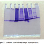 |
Figure 1: Different protein bands on gel electrophoresis |
Acknowledgements
The implementation and success of this project could not have been possible with out the patient guidance of J.MALATHI, Department of Microbiology, Shivani Pharmacy College, Warangal. We are indebted to Sri Dr.Singaracharya, Head of Microbiology Department, Kakatiya University, Warangal for the full fledged co operation. We express our profound gratitude to Principal of Shivani Pharmacy College, for providing excellent lab facility. It’s our privilege to keep on record our deep sense of gratitude and warm regards to all the faculty members for their encouragement
References
- Armstrong AC. Eaton D, E wing JC. Cellular immunotherapy of cancer. Br med J.2001; 323: 1289-1293.
- LH.Hartwell and MB kastan. Cell cycle control and cancer. 1994; 266.
- Anderson W F. gene therapy scores against cancer. nat med 2000; 6: 862-863.
- Frances A shepherd, Srikla S.sridhar. Angiogenisis inhibitors under study for the treatment. Lung cancer.2003; 41:563-572.
- Kathleen Collins, Tylor jacks an Nikola P.pavleich. The cell cycle and cancer PNAS. 1997;34:7
- Kinzler, Kenneth: Vogelstein, Bert,The genetic basis of human cancer. 2002
- Nelson DA, Tan TT, Rabson AB, Anderson D. Hypoxia and defective apoptosis drive genomic stability and tumorigenisis Genes& Development 2004;18(17):2095-107.
- Merlo LM, Pepper JW, Reid BJ, Maley CC. Cancer as an evolutionary and ecological process nat.rev Cancer 6, 2006;(12):924-35.
- Kuper H, Boffetta P, Adami HO. tobacco use and cancer causation association by tumour type, Journal of internal medicine 2006; 252(3): 206-24.
- Sheren keleg, Peter Buchler, Roman Ludwig, Markus w buchler, Helmet frieess,.Invasion and metastasis in pancreatic cancer. Molecular cancer 2003; 2:14.
- Sarah Ahmed saimanaz. Farkhanda ghafoor, M.saleem akhtar. Electrophoretic analysis of serum proteins in prostate cancer. 2004.;43:1
- Sumi S, Arai K,Yoshida K. separation methods applicable to prostate cancer diagnosis and monitoring therapy.J Chromatogr B biomed sci Appl.2001; 764(1-2):445-55.
- Klaus Weber and Mary Osborn. The reliability of molecular weight determinations by dodecyl sulfate poly acrylamide gel electrophoresis. The journal of Biochemistry 1969; 244:16 4406-4412.
- Adjei AA. Blukening of oncogenic radioactive signaling for cancer therapy J.Natl cancer institute .2001; 93:1062-1074.
- Hermans J.F, and Hermans. M-Th Immunoelectrophoresis acta med.Scand 1961; 367; 27-34.
- Muller-Eberhard HJ. A new supporting medium for preparative electrophoresis. Scand J Clin lab., Inv 1960; 12; 33-42.
- Shapiro, viffuella and maizel, Bio chem.. Biophys. Res Commun. 1967; 815.
- G.I. Abelev. Production of embryonal serumα- globulinby hepatomas: review of experimental and clinical data. Cancer research. 1968; 28, 1344-1350.
- John L, Fahey and Alan G.Robinson. Factors controlling serum γ globulin concentration. Published Brit J. 1963; 845-868.
- Cohen S. Gamma globulin metabolism, Brit. med. Bull 1963 in press.
- Olhagen, B. Birke, G.Liijehal, S.O., and Plantin L.O. I131 gamma globulin metabolism in hypergammaglobulinemic states, in proceedings of the XI colloquim on protides of Biological fluids (hub peters editor) amestordam, Elsevier publishing co., Inc., 1963 in press.
- Humphrey, J.H, and Fahey, J.L. The metabolism of normal plasma proteins and γ myeloma protein in mice bearing plasm cell tumours. J.clin, inv.1961; 40: 1696.
- Potter.M, and Kuff,E. Myeloma globulins of plasma cell neoplasms in inbred mice, I. Immunoelectrophorsis of serum, using rabbit antibodies prepared against microsome fractions of the neoplasm, J.Nat. Cancer institute. 1961; 26:1109.
- Askonas, B.A., and Fahey, J.L. An investigation of closely related gamma myeloma proteins and normal mouse gamma globulin by partial enzymatic degradation and starch gel electrophoresis, Nature, 1961; 190:980.

