
Manuscript accepted on :June 29, 2009
Published online on: 13-11-2015
Plagiarism Check: Yes
Vivek Shrivastava and U. K. Jain*
Bhopal Institute of Technology and Science-Pharmacy, Bhojpur road, Bhopal India.
Corresponding Author E-mail:ukjain65@gmail.com
Abstract
The isolation and enrichment of dendritic cell is difficult and time consuming. The complexity in harvesting large number of cells, short survival time, and rapid phenotypic changes in culture have avoided the widespread use of dendritic cells (DC) for fundamental studies Although isolation protocols had been previously established, cells were short-lived and numbers of dendritic cells recovered were less. In present investigation, efforts have been made to modify and improve the method for isolation of dendritic cells. The developed technique improved yield and percentage viability of dendritic cells and up to 62000 dendritic cells can be recovered from one mouse. The technique is easier, quicker, and economic compared to classical method for preparing dendritic cell from skin. In conclusion the generated dendritic cell will likely be useful in a variety of studies related to nanotechnology, allergenicity, microbial infection and transmission, antigen capture and presentation, innate immunity and immunotherapy.
Keywords
Murine cutaneous dendritic cells
Download this article as:| Copy the following to cite this article: Shrivastava V, Jain U. K. An Advanced Method for Isolation of Murine Cutaneous Dendritic Cells and Their Characterization. Biomed. Pharmacol. J.2009;2(1) |
| Copy the following to cite this URL: Shrivastava V, Jain U. K. An Advanced Method for Isolation of Murine Cutaneous Dendritic Cells and Their Characterization. Biomed. Pharmacol. J.2009;2(1). Available from: http://biomedpharmajournal.org/?p=634 |
Introduction
Dendritic cells (DC) were found to be highly effective and superior to other antigen presenting cells when immunizing animals in vivo (Inaba et al., 1998). Methods have been developed recently to grow large number of murine DC from murine bone marrow (Sallusto et al., 1994). For most experimental purposes primary DC, in particular cutaneous DC are needed. The classical approach to obtain murine cutaneous DC consists of culturing epidermal cell suspensions, prepared by trypsinization of epidermis, culturing for three days and subsequently enriching for DC (Schuler et al., 1985), but skin explant cultures are a useful alternative to the trypsinization approach
Materials
RPMI 1640 medium, Trypan blue, Trypsin (2.5%), 2-mercaptoethanol, Ca++ & Mg++ free phosphate buffered saline(PBS) and Ca++ & Mg++ free, Hank’s balanced salt solution (HBSS), were purchased from, HiMedia laboratories Pvt. Ltd. FBS (Fetal Bovine Serum) was from Sigma. C57BL/6 mice of either sex & 6-8 weeks of age were procured from National Institute of Nutrition, Hyderabad and Adenosine –5-triphosphosphoric acid from Otto Kemi.
Methods
Isolation of DC
Isolation was performed by slight modification of procedure described previously by Ortner et. al (Ortner et. al., 1996).Mice were sacrificed with chloroform lethal dose.
A portion of skin 9cm2 (3cm x 3cm) was removed from back of mice which was depleted of hairs previously. All of these were rinsed twice in 70% Ethanol. Ears were cut off and each ear was spliced into two halves using a pair of medium fine forceps (curved and straight forceps) into ventral half (thick) and dorsal half (thin). They were collected on sterile gauze in a petri dish and air dried under hood. Ventral and dorsal Ear halves were placed into separated petridishes. Body wall skin explants were cultured in separate petridishes. The sheets were spread and floated dermal side down. Petridishes were covered with their lids and incubated in CO2 incubator at 37°C (5% CO2). All the tissues were transferred onto culture dishes with fresh medium next day. The cells in the dishes from which tissue had been removed and transferred on day 1 of culture, were cultured until the end of day 2. All cells were pooled at the start of day 3 and were enriched by passing over dense BSA column. Dense BSA column was prepared as described by Schuler (Schuler et al., 1985). DC were identified and enumerated under hemocytometer by their typical ‘veiled’ or ‘hairy’ morphology.
Characterization of dendritic cells
Isolated dermal dendritic cells (DC) were characterized for their membrane ATPase activity. DC in suspension were stained for ATPase using the method of Chaker (Chaker et al., 1984) with minor modifications. A cytocentrifuge preparation of 105 cells was washed with a wash buffer and fixed in winkelman’s fixative. Cells were washed again and covered with a droplet of ATPase reagent. After 45 min incubation in at 370C in a moist chamber, the preparation was washed with wash buffer and treated with dilute ammonium sulphide at room temperature, again washed with water and examined under microscope. Morphological characterization was done by phase contrast microscopy (Figure 1-3) and ultra structural characterization was done by transmission electron microscopy (TEM).(Figure 4)
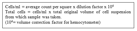
Cell Counting
For cells grown in monolayer cultures, cells were detached from surface of dish using trypsin.Cells were diluted to obtain uniform suspension. Hemocytometer was prepared and filled with diluted cell suspension and slide was viewed on microscope with 100X magnification. Cell counting was performed (Inaba et al., 1998) as follows
Cell Viability
The cell viability was assessed by trypan blue exclusion (Inaba et al., 1998). The cell suspension was diluted 1:10 with trypan blue by adding 10 µL of the cell suspension to 90 µL of trypan blue in a well of a 96-well plate. A Neubauer counting chamber (Feinoptik, Bad Blankanburg, Germany) was filled with the trypan blue-diluted cell suspension. Using an inverted microscope, the number of viable (unstained) cells in the four corner squares was counted. Total cell numbers are counted as following

Results and Discussion
In recent years, the ability of dendritic cells to present tumor-associated antigens is being explored as a possible strategy to boost antitumor immunity in tumor bearing hosts. It has been demonstrated that dendritic cells (DC) derived from lymphoid, nonlymphoid organs and peripheral blood are capable of presenting tumor-associated antigens (Grabbe et al., 1991). With regard to the epidermis, many reports have demonstrated that epidermal langerhans cells are capable of inducing and eliciting antigen specific immunity (Grabbe et al., 1991). Many studies have demonstrated that murine dermis contains antigen-presenting cells, which are capable of processing antigens for the generation of protective immunity in vivo (Grabbe et al., 1991). It is well established that patients immunosuppressed after organ transplantation are at increased risk of developing a variety of malignancies like squamous cell carcinoma, basal cell carcinoma and kaposis’s sarcoma. Thus, immune mechanisms are likely to play a role in the regulation of tumor development in skin. The ability of cutaneous dendritic cells (langerhans cells and dermal dendritic cells) to present tumor associated antigens may be relevant to host defenses against cutaneous malignancies that invade the skin or against metastasis from distant organs that may lodge at this site.
There are two approaches reported to obtain “real dendritic cells” from skin. These are classical method (trypsinization approach) and Skin explant cultures.
The classical method to obtain DC from skin is to enzymatically separate the epidermis from dermis and to digest it into single cell suspension. Typically trypsin is used for this purpose and the resulting suspension contains a low percentage of dendritic cells (1-3%). Several methods are described for efficient enrichment of DC but they are tricky and time consuming and do not yield large numbers of DC. However DC obtained by trypsinization do not represent DC in situ and the mere experimental manipulations like trypsinization, mechanical disruption, centrifugations, etc. are sufficient to irreversibly initiate maturation process. Trypsin also affect cell surface molecules i.e. phenotype.
The migratory capacity is well developed in DC (Ortner et al., 1996), therefore Skin explant cultures are a useful alternative to the trypsinization approach. Skin explant cultures yield populations substantially enriched in DC over a two to four day period. The DC harvested from Skin explant cultures, originate from both dermis and epidermis, i.e., contains both langerhans cells and dermal dendritic cells.
DC were found to be highly effective and superior to other antigen presenting cells when immunizing animals in vivo (Inaba et al., 1998). Larsen et.al (Larsen et al., 1990), developed a method to procure cutaneous migratory dendritic cells, which consists of floating murine whole skin explants (i.e., ear halves) on the standard culture medium so that DC spontaneously emigrate. In Larsen’s procedure skin explants were cultured for a period of three days in the same culture well. The conventional 3 day approach yields an average of 12000 DC per mouse explant (i.e., dorsal ear half). They were enriched to about 40% of all viable cells recovered. Ortner et al. (Ortner et al., 1996) modified this in that they transferred the skin explants to fresh Petri dishes each day, which resulted in higher number of absolute DC and higher percentage of enrichment. The modified approach increases the absolute numbers of DC to three fold and enrichment up to 60 percent. DC were identified and enumerated under hemocytometer by their typical ‘veiled’ or ‘hairy’ morphology.
We used procedure described by Ortner et al. (Ortner et al., 1996) with slight modifications. We modified the procedure in that we used both ear halves i.e., ventral and dorsal to collect migratory DC. Apart from ear, we also used body wall skin to increase the yield of DC per mouse and reduce number of mice. We used keratinocyte-conditioned medium (30% v/v) and we also enriched the DC so obtained by passing them over dense BSA column. At the end of day2 the yield per mouse we got was 62000 and enrichment of 59% (i.e.59% of all viable cells recovered). After passing over dense BSA column enrichment of DC in the low-density fraction was 71 %( i.e.71%of all viable cells recovered) and viability of cells of low-density fraction was more than 90 percent.(Figure 1). This was sufficient enrichment to be used as vaccine, as Koch (Koch et al., 1995) described that even pre-enriched populations (10-15%) are sufficient to perform many studies. While Ortner et. al., used dorsal ear half only and at the end of Day-2, they got 44000 DC per mouse and enrichment of 50%(i.e.50% of all viable cells recovered).The DC harvested from culture medium originate from both dermis and epidermis, i.e., contained both langerhans cells and dermal dendritic cells.
Freshly isolated DC were characterized by (A) Phase contrast microscopy (200x) (B) cytochemical staining of freshly isolated langerhans cells. The ATPase staining procedures are specific for the LC. (Figure 2)
The ATPase reaction is extremely useful tool for scanning langerhans cells with the light microscope and electron microscope. The cell suspension consisted of two clearly different cell types, unstained ones and heavily stained cells (Figure 2). Apart from the different staining properties these two cell populations were morphologically similar with regard to their uniform size and round shape. Both occurred either singly dispersed or in small clusters. The ratio of stained to unstained cells was approximately 1:50.
2-day-old DC were characterized by morphologically by (1) phase contrast microscopy (PCM, Nikon eclipse TS 100) (Figure 3) and (2) Transmission electron microscopy (TEM) .The dendritic nature of DC was shown by phase contrast microscopy. TEM revealed the presence of Birbeck granule, a key ultrastructural feature of DC. (Figure 4).
Conclusions
Skin explant cultures yield populations substantially enriched in DC over a two to four day period. The DC harvested from Skin explant cultures, originate from both dermis and epidermis, i.e., contains both langerhans cells and dermal dendritic cells. The evolved technique is easier, quicker and economic compared to classical method for isolation of dendritic cells from skin.
The generated dendritic cell will likely be useful in a variety of studies related to nanotechnology, allergenicity, microbial infection and transmission, antigen capture and presentation, innate immunity and immunotherapy.
Acknowledgement
We are grateful to Dr. Nikolaus Romani, Professor, Innsbruck University, Austria for providing us technical guidance. We also acknowledge Electron microscopy section of A.I.I.M.S. for performing SEM.
Refrencess
- Inaba, K., Swiggard, W.J., Steinman, R.M., Romani, N., Schuler, G., Isolation of dendritic cells .In current protocols in immunology,J.E.kruisbeek, A.M., Margulies, D.H., Shevach, E.M., and Strober, W., (eds.). Wiley, New York, 3.7.1-3.7.15 (1998).
- Schuler, G., & Steinman, R.M., Murine epidermal langerhans cell mature into potent immunostimulatory dendritic cells in vitro. J.Exp.Med. 161, 526-546 (1985).
- Ortner, U., Inaba, K., Koch, F., Heine, M., Miwa, M., Schuler, G., Romani, N., An improved isolation method for murine migratory cutaneous dendritic cells. Journal Immunological method, 193,71-79(1996)
- Chaker, M.B., Tharp, M.D., Bergstresser, P.R., Rodent epidermal langerhans cells demonstrate greater histochemical specificity for ADP than for ATP and AMP. J.Invest.Dermatol. 82, 496-500 (1984).
- Grabbe, S., Bruvers, S., Gallo, R.L., Knisley, T.L., Nazareno, R., Tumor antigen presentation by murine epidermal cells. Immunol. 146, 3656-3661 (1991).
- Larsen, C.P., Steinman, R.M., Migration and maturation of langerhans cells in skin transplants and explants. J.Exp.Med. 172, 1483-1493 (1990).\
- Inaba, K., Swiggard, W.J., Steinman, R.M., Romani, N., Schuler, G., Isolation of dendritic cells .In current protocols in immunology,J.E.kruisbeek, A.M., Margulies, D.H., Shevach, E.M., and Strober, W., (eds.). Wiley, New York, 3.7.1-3.7.15 (1998).
- Koch, F., Trockenbacher, B., kampgen, E., Antigen processing in populations of mature murine dendritic cells is caused by subsets of incompletely matured cells. Immunol. 155, 93-100 (1995).

