
Manuscript accepted on :01-Nov-2018
Published online on: 11-12-2018
Plagiarism Check: Yes
Reviewed by: Saranya V
Second Review by: Abdalbasit Al-Ubaidi
Final Approval by: Prof. Juei-Tang Cheng
Farah Salahalden Abbas , Nadeen Jamal Abdulredah and Amer Salman Hassan
Department of Conservative Dentistry, Al Mustansiriyah Dental College, Baghdad, Iraq.
Corresponding Author E-mail: haneenmustafa292@ yahoo.com
DOI : https://dx.doi.org/10.13005/bpj/1596
Abstract
Endodontic therapy is essentially a debridement procedure that requires the removal of the irritants of the canal and periapical tissue if success is to be gained. The debridement may include instrumentation of the canal, placement of medicament and irrigants. Complete cleaning of the root-canal system requires the use of irrigants that dissolve organic and inorganic material. The study aimed to evaluate changes in dentin microhardness after canal irrigation with different solutions. Twenty four freshly extracted human mandibular molars (distal roots with single canals) were used. 10mm root length was taken as standard length. The roots were embedded into auto polymerizing acrylic resin using plastic molds before the canals preparation and micro hardness test. The distal roots were prepared with one shape rotary file. Before the preparation each root was irrigated with 1ml distilled water. Then the roots were divided into four groups according to the final irrigation protocol: Group A: NaOCl 2.5%, Group B: EDTA 17%, Group C: Citric Acid 40%, Group D: Distilled Water. For (Vicker microhardness test) the same load and time 500 g test load for 20 seconds, will be conducted three times at distance 0.5mm from canal lumen ; thus there will be 9 indentations on each specimen surface. An average of the three readings for each test condition will be recorded as the VHN value of a specimen. Comparing all four groups statistically there was no significant difference among them. The mean values were found more reduced in EDTA group followed by NaOCL group, and then Control and Citric Acid groups. All the groups showed reduction in dentin microhardness. EDTA group showed the maximum reduction followed by NaOCL group, and least with Citric Acid group.
Keywords
Citric acid; EDTA; Irrigation; Microhardness; Vicker test
Download this article as:| Copy the following to cite this article: Abbas F. S, Abdulredah N. J, Hassan A. S. Effect of Final Irrigation Protocol on Dentin Microhardness. Biomed Pharmacol J 2018;11(4). |
| Copy the following to cite this URL: Abbas F. S, Abdulredah N. J, Hassan A. S. Effect of Final Irrigation Protocol on Dentin Microhardness. Biomed Pharmacol J 2018;11(4). Available from: http://biomedpharmajournal.org/?p=24610 |
Introduction
Endodontic therapy is essentially a debridement procedure that requires the removal of the irritants of the canal and periapical tissue if success is to be gained. The debridement may be carried out in various ways as the case demands and may include instrumentation of the canal, placement of medicament and irrigants.1 The main goal of instrumentation is to facilitate effective irrigation, disinfection, and filling. Several studies using advanced techniques such as micro computed tomography (CT) scanning have demonstrated that proportionally large areas of the main root-canal wall remain untouched by the instruments,1 emphasizing the importance of chemical means of cleaning and disinfecting all areas of the root canal.2
Microhardness defined as the resistance to local deformation and it tests based on the induced permanent surface deformation that remains after removal of load. Any change in the microhardness of the root dentin may adversely affect sealing ability and adhesion of dental material such as resin cements and root canal sealers to dentin.
Microhardness tests are commonly used to study the physical properties of materials, and they are widely used to measure the hardness of teeth. 3,4,5 This method is easy, quick, and requires only a tiny area of specimen surface for testing. Using this technique, the specimen surfaces were impressed with a diamond indenter (a Knoop or a Vickers) at a certain load for a certain period of time. After load removal, diagonals of the indentation were measured with an optical microscope. The hardness number was defined by the ratio between the indentation load and the area of the residual impression, which depended on the indenter shape.
There is no single irrigating solution that alone sufficiently covers all of the functions required from an irrigant. Optimal irrigation is based on the combined use of 2 or several irrigating solutions. Complete cleaning of the root-canal system requires the use of irrigants that dissolve organic and inorganic material.2
Sodium hypochlorite is the most popular irrigating solution and is commonly used in concentrations between 0.5% and 6%. It is a potent antimicrobial agent, killing most bacteria instantly on direct contact. It also effectively dissolves pulpal remnants and collagen, the main organic components of dentin.Although hypochlorite alone does not remove the smear layer, it affects the organic part of the smear layer, making its complete removal possible by subsequent irrigation with EDTA or citric acid. The presence of inactivating substances such as exudate from the periapical area, pulp tissue, dentin collagen, and microbial biomass counteract the effectiveness of NaOCl.6
As hypochlorite is active only against the organic matter, other substances must be used to complete the removal of the smear layer and dentin debris. EDTA and Citric Acid effectively dissolve inorganic material, including hydroxyapatite7,8 they have little or no effect on organic tissue and alone they do not have antibacterial activity.
Aim of the study
The study aimed to evaluate changes in dentin microhardness after canal irrigation with different solutions.
Material and Methods
Sample selection
Twenty four freshly extracted human mandibular molars (distal roots with single canals) were used.
Selection criteria
Straight roots with single canals.
Free from caries and cracks.
Centered apical foramen.
Roots without resorption.
Roots length 11mm.
Sample preparation
The teeth were cleaned of all debris and stored in distilled water till the time of preparation. Teeth were sectioned transversely at cemento- enamel junction using diamond disc operated by low speed hand piece under continuous water coolant. Remnant of pulp tissue were removed by barbed broaches, then the patency of the canals were determined with S.S. k-file size 10,15 until it was visible at apical foramen and the working length were established 10 mm. The roots were embedded into auto polymerizing acrylic resin using plastic molds before the canals preparation and micro hardness test.
Root canal preparation
The distal roots were prepared with one shape rotary file operated by x-smart micro motor at speed 350-450 r.p.m., torque 1.5 N/cm, gear ratio 16:1. Before the preparation each root was irrigated with 1ml distilled water and one shape rotary file was operated in the canal not more than 1 minute, then the specimens were irrigated with 5 ml of each test solution to receive the final irrigation according to the sample grouping.
Sample grouping
The roots were divided into four groups according to the final irrigation protocol:
Group A: 6 roots were irrigated with 5ml NaOCl 2.5%.
Group B: 6 roots were irrigated with 5ml EDTA 17%.
Group C: 6 roots were irrigated with 5ml Citric Acid 40%.
Group D: 6 roots were irrigated with 5ml Distilled Water.
Microhardness Test
For microhardness test the specimens were ground flat on a circular grinding machine with ascending grades of SiC abrasive papers (400 and 1000 grit) under constant water irrigation on rotary felt disk. Then each test condition with the same load and time will be conducted three times at distance 0.5mm from canal lumen as shown in figure (1) using Vicker Micro hardness Machine as shown in figure (2); thus there will be 9 indentations on each specimen surface obtained from 500 g test load for 20 seconds. An average of the three readings for each test condition will be recorded as the VHN value of a specimen.
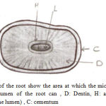 |
Figure 1: Cross section of the root show the area at which the microhardness of root canal dentin measured., L: Lumen of the root can , D: Dentin, H: area which the hardness measured(0.5mm from the lumen) , C: cementum. |
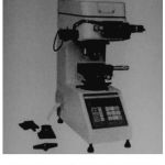 |
Figure 2: Vickers Micro hardness Machine
|
Results
The results of Descriptive Statistics which include mean, standard deviation, standard of error, minimum and maximum for all groups are shown in Table (1).
Table 1: Descriptive statistics
| N | Mean | Std. Deviation | Std. Error | 95% Confidence Interval for
Mean |
Minimum | Maximum | ||
| Lower Bound | Upper Bound | |||||||
| NaOCL
EDTA Citric Acid CONTROL Total |
6
6 6 6 24 |
48.300
43.000 58.133 54.567 51.000 |
2.3281
3.2533 3.9343 3.1379 6.6464 |
.9504
1.3282 1.6062 1.2811 1.3567 |
45.857
39.586 54.005 51.274 48.193 |
50.743
46.414 62.262 57.860 53.807 |
45.9
40.0 54.3 50.4 40.0 |
52.1
47.3 62.5 59.3 62.5 |
It has shown that EDTA group has lowest mean values of dentin microhardness after final irrigation protocol. And Citric Acid group has the highest mean values of dentin microhardness.
Analysis of variance ANOVA test was performed to identify the presence of any statistically significant difference among the means of microhardness reduction for all groups Table (2).
Table 2: Analysis of variance test (ANOVA)
| Sum of Squares | df | Mean Square | F | Sig. | |
| Between Groups
Within Groups Total |
809.373
206.647 1016.020 |
3
20 23 |
269.791
10.332 |
26.111 | .000 |
ANOVA test revealed that there was no statistically significant difference (P ˃ 0.05) among the groups.
The Least signifigant different test (LSD) was performed for multiple comparisons between groups Table (3).
Table 3: Least signifigant different test (LSD)
| (I) Irrigation Solution | (J) Irrigation Solution | Mean
Difference (I-J) |
Std. Error | Sig. | 95% Confidence Interval | |
| Lower Bound | Upper Bound | |||||
| NaOCL | EDTA
Citric Acid CONTROL |
5.3000*
-9.8333* -6.2667* |
1.8558
1.8558 1.8558 |
.010
.000 .003 |
1.429
-13.705 -10.138 |
9.171 -5.962
-2.395 |
| EDTA | NaOCL
Citric Acid CONTROL |
-5.3000*
-15.1333* -11.5667* |
1.8558
1.8558 1.8558 |
.010
.000 .000 |
-9.171
-19.005 -15.438 |
-1.429
-11.262 -7.695 |
| Citric Acid | NaOCL EDTA
CONTROL |
9.8333*
15.1333* 3.5667 |
1.8558
1.8558 1.8558 |
.000
.000 .069 |
5.962
11.262 -.305 |
13.705
19.005 7.438 |
| CONTROL | NaOCL
EDTA Citric Acid |
6.2667*
11.5667* -3.5667 |
1.8558
1.8558 1.8558 |
.003
.000 .069 |
2.395
7.695 -7.438 |
10.138
15.438 .305 |
The results of (LSD) test showed that there was a significant difference between NaOCL and EDTA groups and there was a significant difference between NaOCL and and control groups.
Discussion
Irrigation is presently the best method for the removal of tissue remnants and dentin debris during instrumentation. Numerous solutions have been recommended for use as root canal irrigants.1 The effect of mechanical washing, reduction of friction, and control of temperature are all important underlying reasons for irrigation; however, the most important tasks are dissolution of organic and inorganic tissue, and killing of the microbes.
The study aimed to evaluate changes in dentin microhardness after canal irrigation with different solutions; and in this study we use three of commercially available irrigating solutions. Research and clinical experiences have shown that NaOCl has several properties that contribute to effective chemomechanical debridement of a root canal system. The use of chelating agents (EDTA and Citric Acid) for final irrigation removes the smear layer and reduces dentin microhardness, which increases the access of the irrigant to dentinal tubules, allowing for proper disinfection.9
In the present study EDTA promoted the largest reduction in dentin microhardness at 0.5 mm from canal lumen. These results are in agreement with those of several previous studies 9,10,11,12,13 in which this solution also reduced microhardness. This effect is desirable in the layer next to the canal lumen and it has been associated with increasing calcium loss, resulting in dentin demineralization and softening.
The use of 2.5% Sodium Hypochlorite as a root canal irrigation significantly reduce the microhardness of root dentin this due to organic dissolving properties of Sodium Hypochlorite on collagen component of dentin.14 In addition to that Sodium Hypochlorite extract the Calcium ion from the dentin and decrease the calcium/ phosphorus ratio.15,16,17 The current study agree with the study by Slutzky-Goldberg et al,18 Ari et al19 and Oliveira et al20 who conclude that Sodium Hypochlorite significantly reduces the microhardness of root canal dentin.
In the present study Citric Acid group did not significantly change microhardness at 0.5 mm from canal lumen. The results of control group and citric acid group showed that there was no statistically significant difference between them, and these results may be related to that the teeth have different initial physical characteristics10,21,22 and the initial microhardness of root dentin in this study not evaluated.
The primary factors that govern the action of an irrigant are the contact time and the concentration; and in the present study we used the root canal irritant for 5 minutes in our microhardness test and this is in agreement with studies Ulusoy & Görgül23 and Sayin et al16 who use the root canal irrigants in their microhardness tests for 5 minutes, stating that this duration is more realistic in terms of clinical practice. The solutions were taken to the canal with the help of a syringe coupled to the irrigation needle, thus simulating clinical practice.
Calt & Surper, 24 their study suggested that one minute application of 17% EDTA was effective to remove the smear layer. But previous studies evaluated the effect of root canal irrigants on the microhardness of the root canal dentin for five minutes.23,25 Likewise, in the present study we use test solutions for 5 minutes.
Another determinant that has a profound effect on the post-treatment microhardness values of dentin is the concentration of the irrigating solution.25 As the concentration of NaOCl increases, its bactericidal and smear layer removal efficacy also increases (26, 27). Most studies showed that 17% EDTA was effective to remove the smear layer but a few reports have indicated that solutions with lower concentrations (eg, 10%, 5%, and even 1%) remove the smear layer equally well after NaOCl irrigation. Citric Acid is also marketed and used in various concentrations, ranging from 1% to 50%, with a 10% solution being the most common.
Conclusions
All the groups showed reduction in dentin microhardness. EDTA group showed the maximum reduction followed by NaOCL group, and least with Citric Acid group.
References
- Weine F. S. Endodontic therapy, Sixth Edition, Mosby. 2004;4.
- Peters O. A., Scho¨nenberger K., Laib A. Effects of four Ni-Ti preparation techniques on root canal geometry assessed by micro computed tomography. Int Endod J. 2001;34:221–30.
CrossRef - Chunmuang S., Jitpukdeebodintra S., Chuenarrom C and Benjakul P. Effect of xylitol and fluoride on enamel erosion in vitro. Journal of Oral Science. 2007;49(4):293-297.
CrossRef - Faraoni-Romano J. J., Turssi C. P and Serra M. C. Concentration-dependent effect of bleaching agents on microhardness and roughness of enamel and dentin. American Journal of Dentistry. 2007;20(1):31-34.
- Attin T., Meyer K., Hellwig E., Buchalla W and Lennon A. M. Effect of mineral supplements to citric acid on enamel erosion. Archives of Oral Biology. 2003;48(11):753-759.
CrossRef - Haapasalo H. K., Siren E. K., Waltimo T. M. Inactivation of local root canal medicaments by dentine: an in vitro study. Int Endod J. 2000;33:126–31.
CrossRef - Loel D. A. Use of acid cleanser in end odontic therapy. J Am Dent Assoc. 1975;90:148–51.
CrossRef - Haapasalo M., Ørstavik D. In vitro infection and disinfection of dentinal tubules. J Dent Res. 1987;66:1375–9.
CrossRef - Saghiri M. A., Delvarani A., Mehrvarzfar P., Malganji G., Lotfi M., Dadresanfar B. A study of the relation between erosion and microhardness of root canal dentin. Oral Surg Oral Med Oral Pathol Oral Radiol Endod. 2009;108(6):e29-34.
CrossRef - Taneja S., Kumari M., Anand S. Effect of QMix, peracetic acid and ethylene diamine tetra acetic acid on calcium loss and microhardness of root dentine. J Conserv Dent. 2014;17(2):155-8.
CrossRef - Tartari T., Souza P. A. R. S., Almeida B. V. N., Junior J. O. C. S., Pessoa O. F., Junior M. H. S. S. A new weak chelator in endodontics: effects of different irrigation regimens with etidronate on root dentin microhardness. Int J Dent. 2013;2013. ID 743018.
- Aranda-Garcia A. J., Kuga M. C., Chavéz-Andrade G. M., Kalatzis-Sousa N. G., Hungaro Duarte M. A., Faria G. Effect of final irrigation protocols on microhardness and erosion of root canal dentin. Microsc Res Tech. 2013;76(10):1079-83.
CrossRef - Das A., Kottoor J., Mathew J., Kumar S., George S. Dentine microhardness changes following conventional and alternate irrigation regimens an in vitro study. J Conserv Dent. 2014;17(6):546-9.
CrossRef - Farag A., Hassanien E. Effect of Chemical irrigation and Intracanal CO2 Laser Irradi-ation on Hardness of Root Canal Dentin. Cairo Dent J. 2000;16:135-139.
- Dagan H., Calt S. Effect of Chelating Agents and Sodium Hypochlorite on Mineral Content of Root Dentin. J Endod. 2001;9:578-580.
CrossRef - Sayin C., Cehreli Z., Deniz D., Akcay A., Tuncel B., Dagli F., Gozukara H and Ka-layci S. Time-dependent Decalcifying Effects of Endodontic Irrigants with Anti-bacterial Properties. J Endod. 2009;35:280-283.
CrossRef - Gurbuz T. Evaluation of Root Canal Dentin after ND:YAG Laser Irradiation and Treated with Five Different Irrigation Solutions: A Preiminary Study. J Endod. 2009;34:318-321.
CrossRef - Slutzky- Goldberg I., Maree M., Liberman R and Heling I. Effect of Sodium Hypochlorite on Dentin Microhardness. J Endod. 2004;30:880-882.
CrossRef - Ari H., Erdemir A and Belli S. Evaluation of the Effect of Endodontic Irrigation Solution on the Microhardness and Roughness of Root Canal Dentin. J Endod. 2004;30:792-797.
CrossRef - Oliveira L., Carvahlo C., Nunes W., Valera M., Camargo C and Jorge A. Effect of Chlorhexidine and Sodium Hypochlorite of Root Canal Dentin. Oral Surg Oral Med Oral Pathol Oral Radiol Endod. 2007;104:125-133.
CrossRef - Cruz-Filho A. M., Sousa-Neto M. D., Savioli R. N., Silva R. G., Vansan L. P., Pécora J. D. Effect of chelating solutions on the microhardness of root canal lumen dentin. J Endod. 2011;37(3):358-62.
CrossRef - Ghisi A. C., Kopper P. M., Baldasso F. E., Stürmer C. P., Rossi-Fedele G., Steier L et al. Effect of super-oxidized water, sodium hypochlorite and EDTA on dentin microhardness. Braz Dent J. 2014;25(5):420-4.
CrossRef - Ulusoy Ö. İ., Görgül G. Effects of different irrigation solutions on root dentine microhardness smear layer removal and erosion. Aust Endod J. 2013;39:66–72.
CrossRef - Calt S., Serper A. Time-dependent effects of EDTA on dentine structures. J Endod. 2002;28:17–9.
CrossRef - Zhang K., Kim Y. K., Cadenaro M., Bryan T. E., Sidow S. J., Loushine R. J. Effects of different exposure times and concentrations of sodium hypochlorite /ethylenediaminetetraacetic acid on the structural integrity of mineralized dentin. J Endod. 2010;36:105–9.
CrossRef - Zehnder M. Root canal irrigants. J Endod. 2006;32:389–98.
CrossRef - Marending M., Luder H. U., Brunner T. J., et al. Effect of sodium hypochlorite on human root dentine–mechanical, chemical and structural evaluation. Int Endod J. 2007;40:786–93.
CrossRef

