
Manuscript accepted on :16 May 2018
Published online on: 08-06-2018
Plagiarism Check: Yes
Fahmida binti Abd Rahman1, Deepa Gurunathan2 and Madhu Sudhan Vasantharajan3
1Paediatric dentistry, Saveetha Dental College and Hospital, Chennai, India.
2Department of Pedodontics Saveetha Dental College, Saveetha University 162, Poonamalle High Road Chennai 600077, Tamil Nadu, India.
3Pediatric dentistry, Saveetha Dental College and Hospital, Ponamalle high road, Chennai-600127, India.
Corresponding Author E-mail : drgdeepa@yahoo.co.in
DOI : https://dx.doi.org/10.13005/bpj/1475
Abstract
Radiography in dentistry is commonly used in modern dental health care. It acts as a diagnostic tool in identifying the physical condition of the patients. However the use of dental radiography should be carefully managed since it can cause some side effects toward normal cells and tissues especially in growing children. To assess the knowledge, attitude and practice of undergraduate dental students on radiation exposure protection for pedodontic patients. A self-administered questionnaire consists of 13 questions with both ‘yes’,’no’ and multiple choice pattern was prepared to obtain information about knowledge, attitude and practice on radiation exposure protection for pedodontic patients.A total of 100 dental students were chosen as the participants. The questionnaires were assessed by manual survey. The use of dental radiograph has bee significantly enhanced over the years for better diagnosis and treatment planning .Tough the application of has been increased, there is an increased risk for unwanted exposure for both patient and operator also. In this current study, conducted among third year and final year dental students, we observed that the students are able to take proper radiograph with unnecessary retakes with years of study and practice. To conclude, it can be noted that the final year dental students were much more knowledgeable in using radiograph judicially compared to the other group of participants which belong to third year dental students. However, in terms of practise both third year and final year dental students irrespective of year of study do not follow radiation protection measures even though they were aware of it.
Keywords
Attitude; Dental Radiograph; Exposure; Retakes; Side Effects
Download this article as:| Copy the following to cite this article: Rahman F. B. A, Gurunathan D, Vasantharajan M. S. Knowledge, Attitude and Practice of Radiation Exposure Protection for Pediatric Patients Among Undergraduate Dental Students. Biomed Pharmacol J 2018;11(2). |
| Copy the following to cite this URL: Rahman F. B. A, Gurunathan D, Vasantharajan M. S. Knowledge, Attitude and Practice of Radiation Exposure Protection for Pediatric Patients Among Undergraduate Dental Students. Biomed Pharmacol J 2018;11(2). Available from: http://biomedpharmajournal.org/?p=20636 |
Introduction
Radiation can be refers to the movement of energy though space and matter. It can occur either in particulate or in electromagnetic radiation. As the name implies, electromagnetic radiation is the movement of energy through space with the joining of electric and magnetic field. When the velocity of charged particle changes, the electromagnetic radiation can be established. Examples of electromagnetic radiation are x rays, y rays, uv rays, visible light, infrared radiation, microwaves and radio waves. Radiation types for electromagnetic radiation depend on their energy. It can be available as ionizing or nonionizing radiation.1-5 Ionizing radiation is quiet harmful to cells of human body as the fact that it can causes numerous side effects toward biological with the establishment of free radicals. It can give side effects either directly or indirectly which can cause DNA damage, involving breaking of single or double strand and DNA cross links. Based on the history of radiology, x rays were discovered by Professor William Conrad Roentgen from Germany. He successfully discovered the rays after attempting repeated investigations using cathode ray tube in his laboratory. Dr. C. Edmund Kells was the first to use dental radiograph in United States [6]. Radiology plays a significant role in the oral health care of infants, children, adolescent and elder patients. In dentistry, radiograph is mainly used to diagnose and evaluate problem related to oral diseases and for a better treatment planning. Radiology has grown rapidly with the advancement and expansion of imaging facilities such as Cone Beam Computed Tomography (CBCT), Computed Tomography (CT) and ortho cubic super high resolution CT (Ortho CT). Based on statistics, it is recorded that approximately 480 million dental radiographic examination has been done per year accounting for about 15 % of total diagnostic x ray examination.7 High radiation exposure towards pediatric patients can lead to biological effects that can last for a long period of time.8 It is also believed that pediatric patients are more prone to develop cancer for the same dose received for the adults.9 Biological effects can be divided into two sub classes which are non-stochastic (deterministic effects) and stochastic effects. The severity of the response is directly proportional to the amount or dose of radiation. It is a dose dependent which cause biological damage in human body. On the other hand, stochastic effect is non dose dependent.
Deterministic effects and stochastic effects will be only achieved through high dose ionising radiation or known as X rays. If low dose of radiation is use, it may lead to stochastic effects. Even though the dose of radiation used during radiographic examination is in a low dose but still it is paramount to reduce radiation exposure toward our body as accumulated dose may happen. According to this, dental radiograph should be only taken when it is really necessary and should be avoided as much as possible if the risks outweighs its benefit. Guidelines for restrictions on the amount of radiation perceived by both dentist and patients has bene introduced by National Council on Radiation Protection and Measurement (NRCP) and the International Commission Council on Radiological Protection (ICRP) when they recognize harmful effects of radiation and the associated risks. The first guideline in radiation protection is the justification principle. This principle states that dental radiographic examination can be done on patients only when the benefits outweigh the risk of radiation exposed. Other than that, the second principle is the optimization principle. This principle state that dentists should use every means or effort to lower or eliminate unnecessary exposure towards their patients. The third principle is known as ALARA principle. ALARA is the short term for “as low as reasonably achievable”.10 A dentist should develop professional judgement after thorough history taking and clinical examination. All three principles should be keep in mind before proceed the patients for radiographic examination.11 Currently, newer technologies have been established such as the utilize of digital radiography which is rapidly enhancing and now coming to the fore. Some of the benefits of use of using digital radiography is that it can be used for referral purposes, distinct environmental advantages and most paramount is that it may lower radiation exposure,12,13 In addition, effort like use of lead apron, thyroid collar, high speed film and proper angulation during taking radiograph should be taken seriously in order to reduce the risk of radiation exposure towards pediatric patients.14 The principle of radiation protection is not only to reduce exposure of radiation towards pediatric patients but it should also provide positive outcome from the use of dental radiograph.15 Negligence toward radiation exposure protection may lead to unnecessary radiation effect. Most of the cases in dental clinic require dental radiograph examination for better and clear diagnosis of the disease. During clinical rotation, all of the undergraduate and postgraduate training dentists are trained to carry out the dental radiograph examination by themselves under supervision. Commonly, undergraduate dental students training will be divided into pre clinical level and clinical level. Preclinical level students comprise of first and second year students whereas clinical students comprise of third, fourth and interns . Most of the previous studies have shown that there was poor knowledge among dental students,16 dentists,17 and other health workers.18 The radiation protection knowledge and practice of dental radiography by dentists is consequently important. However, the proper radiation protection practice by dentists is still insufficient. Third year, final year dental students and intern should have better and thorough knowledge toward biological hazards of x rays and different protection protocols as they are the one that will have high risk of getting drawback of radiation exposure as they cannot avoid themselves from using x ray machine in their practises. In comparison to the dose of radiation exposure in medical course, the radiation dose apply in dental imaging is significantly low but due to repeated examinations over time it can produces high cumulative doses. Therefore, this current study was conducted to assess knowledge, attitude and practise of radiation exposure protection for paediatric patients among undergraduate dental students in a private dental college in Chennai.
Method and Materials
A self administered questionnaires comprises of 12 questions with ‘yes’ and ‘no’ pattern and multiple choice question was prepared and piloting was done to obtain information about knowledge, attitude and practice radiation. The questionnaire was developed in English language. A total of 200 undergraduate dental students were chosen as the participants. Study objectives were explained to the participants and it was distributed in a class room setting and the responses from all students were collected as soon as they filed up the questionnaires which was on the same visit. The questionnaires were distributed manually to them. After collecting data,descriptive statistical analysis was performed.Table 1 shows the result regarding respective questions. According to the question on awareness of need of peadiatric radiographs, it is seenthat the majority of the final year students (80%) aware regarding the purpose of dental radiographs for pediatrics whereas the level of awareness for 3rd year students still low which has percentage of 70 %.
The result shows that almost all third year and final year students have been adequately trained to diagnose pedodontic patients. Both third and final years are aware that the Xray radiation is harmful for growing children.
Table 1
| No of question | 3rd year dental students | 4th year dental students |
| 1.Have you been trained to diagnose pedodontic patients ?
a) Yes b)No |
80(80%)
20(20%) |
100(100%)
0 |
| 2. Are you aware of pediatric dental radiograph for ?
a) Yes b) No |
70(70%)
30(30%) |
80(80%)
20(20%) |
| 3. Are you aware of radiation protection options available for children ?
a) Yes b)No |
50(50%)
50(50%) |
84(84%)
16(16%) |
| 4. Can x rays be reflected from the body of the subject ?
a) Yes b)No |
76(76%)
24(24%) |
82(82%)
18(18%) |
| 5. Are you aware about the distance for both
operator and children during during dental radiographic examination? a)Yes b)No |
22(22%)
78(78%) |
30(30%)
70(70%) |
| 6.Do you agree that dental x-rays are harmful for a growing child?
a) Yes b) No
|
100(100%) 0 |
100(100%) 0 |
| 7.Do you regularly prefer using lead aprons?
a)Yes b) No |
22(22%)
78(78%) |
30(30%)
70(70%) |
| 8.Which cells are more vulnerable for radiation?
a)thyroid gland b)growing bone c)salivary gland d)optic lens |
34(34%)
18(18%) 36(36%) 12(12%) |
60(60%)
10(10%) 14(14%) 16(16%) |
| 9.What film you will use for radiation?
a) E speed b) F speed |
36(36%)
64(64%) |
60(60%)
40(40%) |
Results
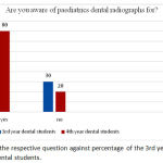 |
Figure 1: shows the respective question against percentage of the 3rd year and final year dental students.
|
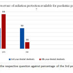 |
Figure 2: shows the respective question against percentage of the 3rd year and final year dental students.
|
The result shows a percentage of 84 % final year students aware about radiation protection options available for children meanwhile only 50 % percentage of third year students aware about it.
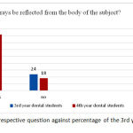 |
Figure 3: shows the respective question against percentage of the 3rd year and final year dental students.
|
The results from the participants revealed that a percentage of 76% of third year dental students agreed that x ray can be reflected from the body of the subject whereas 4th year dental students had a percentage of 82% which is a little higher than 3rd year students.
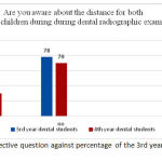 |
Figure 4: shows the respective question against percentage of the 3rd year and final year dental students.
|
The data shows that most of the students do not aware the position and distance rule and thus are at a risk of unnecessary risk of radiation exposure.
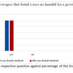 |
Figure 5: shows the respective question against percentage of the 3rd year and final year dental students.
|
The results from the participants revealed that all of them strongly agree that dental x rays are harmful for a growing child.
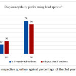 |
Figure 6: shows the respective question against percentage of the 3rd year and final year dental students.
|
It was observed that both categories of dental students lack of awareness regarding the importance of wearing lead aprons during radiographic examination.
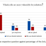 |
Figure 7: shows the respective question against percentage of the 3rd year and final year dental students.
|
The response for this question among the participants has been primarily among thyroid gland and salivary gland in the beginning but participants of final year had given clearly thyroid gland as the organ that must be protected during radiation exposure.
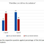 |
Figure 8: shows the respective question against percentage of the 3rd year and final year dental students
|
The response received from these participants showed that both e and f speed film are commonly used by both 3r year and final year students.
Discussion
The use of dental radiograph has bee significantly enhanced over the years for better diagnosis and treatment planning.19 Tough the application of has been increased, there is an increased risk for unwanted exposure for both patient and operator also. Many factors such as proper technique, positioning and equipment play major role in the radiographic protection for operator and patients especially paediatric patients.20 In this current study, conducted among third year and final year dental students, we observed that the students are able to take proper radiograph with unnecessary retakes with years of study and practice. This kind of result can be correlates with previous studies carried out in Turkey,21 Belgium22 and Canada21 The results for this study showed that participants are well aware of the main role of imaging science. In addition, level of knowledge regarding radiation exposure protection available also increase corresponding to their level of academic. This is similar to the result obtained by a study done in Ukraine. In our study, most of the third year students were aware of the purpose of taking radiation for pediatrics but this knowledge was comparatively low in the graduates compared to final year dental students. When third year and final year dental students have been asked regarding the important organ need to be kept away or protect from dental radiography a total of 94% participants were aware that thyroid is the most sensitive organ towards radiation compare to any other organ followed by salivary glands as the second organ. This is same with the result from Ukraine study in which 66% of the participants answered thyroid as the important organ to be protected.23 From our current study, it can be noted that the final year students being more aware that X rays can be reflected from body of object compared to third year dental students. One of the significant findings in this study is that there was less number of students using lead apron during operating an x ray unit considering the positive effect of lead apron. The percentage for third year students that always wore lead apron was 22% while the percentage for final year students that wore lead apron was 30 % which can be considered as critical condition. This finding can be compared with the study carries out by R. Jacobs and his associates in between the Belgian dentists where only 12 % of dentists wore lead apron during radiographic examination. In our opinion, the fact that may urge them not to wore lead apron among third year and final year students maybe because of non-availability of lead apron or less availability. Other than that, the reason why they did to wore lead apron maybe because of the increased weight of the apron itself. Knowledge of the students regarding type of film used for radiation examination is better. One of the striking finding of our study was the reduced number of 3rd year students wearing lead apron while operating an x-ray unit, considering the beneficial effect of lead apron. In our present study, the percentage of intern participants that always wore lead apron was 84.61% which was comparatively better as compared to the study carried out by R. Jacobs and his associates amongst the Belgian dentists where only 12% of the dentists wore lead apron while operating an x-ray units.23 The reasons for not wearing a lead apron among undergraduates might be attributed to the non-availability of lead apron and increased weight of the apron.
From our current study, it is undeniable that the participants were aware of the radiation exposure protection available for pediatric patients and it is more so with the final year and then followed by the third year dental students. Eventhough there were so not many studies were available for comparison, the results fits well with our hypotheses that the KAP of undergraduates towards radiation protection was limited and this can be applicable to this community as well. Further studies with a larger sample size are needed to validate our hypotheses.24 More over the current study was a single institutional based one, hence a cross- sectional study comprising of similar samples utilizing multiple institutional participants are essential for authentication.There are many ways or management options available for radiation exposure protection for both paediatric patients and operator. These factors include proper angulation, proper distance between x ray machine and operator, proper technique used, proper equipment used for patients, suitable speed and type of film. Apart from that, training of personnel in radiation protection should be a continuous process rather than just only when the patients had patients that required x ray for better diagnosis and treatment planning even after graduation from dental school to achieve long- term knowledge retention and repeated reinforcements. It is also advised that radiographic examinations at the undergraduate level should be taken intricately so as to inculcate knowledge about radiation hazards as early as possible. After graduation, the practitioners should attend dental education programs continuously to increase and keep their knowledge up to date. This can help in keeping the dentist updated with any new information for his practice. In addition, awareness regarding radiation exposure hazards should be circulated on social media to reach a larger audience. The periodic check-up of X-ray units should also be made as mandatory by the authorities. Furthermore, film badges for personal dosimetry should be made compulsory to be worn by the dentist and the para-medical staff to keep an eye on the amount of radiation exposure.
Conclusion
This current study was conducted in a Dental institution with intra oral and extra oral dental radiographic facilities. Based on the result of this study ,it can be noted that the final year dental students were much more knowledgeable in using radiograph judicially compared to the other group of participants which belong to third year dental students . However, in terms of practise both third year and final year dental students irrespective of year of study do not follow radiation protection measures even though they were aware of it. The side effect of radiation exposure is dangerous to both patients as well as for the dentists. Therefore, it is necessary to reduce the exposure towards dental radiation.
Acknowledgement
The author(s) received no specific funding for this work.
References
- Haring J.I, Lind L.J. Chapter 5: Radiation protection in textbook of dental radiography principles and techniques. B. Saunders Company. 1996;64-79.
- Ribeiro D.A, de Oliveira G, de Castro G, Angelieri Cytogenetic biomonitoring in patients exposed to dental X-rays: comparison between adults and children. Dentomaxillofacial Radiol. 2008;37(7): 404-407.
CrossRef - Prabhat M.P.V, Sudhakar S, Kumar B.P. Knowledge, attitude and perception (KAP) of dental undergraduates and interns on radiographic protection- A questionnaire based cross-sectional study. J Adv Oral Research. 2011;2(3):45-49.
CrossRef - Little M.P, Wakeford R, Tawn E.J, Bouffler S.D, Berrington de Gonzalez A. Risks associated with low doses and low dose rates of ionizing radiation: linearity may be (almost) the best we can do. Radiology. 2009;251(1):6-12.
CrossRef - White S.G, Pharoah M.J. Oral radiology: Principles and interpretation. (5th edn), Mosby. St. Louis, USA. 2004;25-46.
- Judit Forrai. History of X Ray In Dentistry. Rev. Clin. Pesq. Odontal. 2007; set/dez;3(3):205-211
- UNSCEAR Report to the General Assembly, with scientific annexes. Sources and effects of ionising radiation. Volume 1: Annex A: Medical Radiation Exposures. Available from: http://www.unscear.org/docs/reports/2008/0986753_Report_2008_Annex_A.pdf. 2008; [Last accessed on 2013 Oct 08].
- Furlow B. Radiation Protection in Pediatric Imaging. Radiologic Technology. 2011;82(5):421-439.
- Karami V, Zabihzadeh M, Gholami M, Shams N, Fazeli-Nezhad Z. Dose reduction to the Thyroid Gland in Pediatric Chest Radiography. Int J Pediatr. 2016;4(5):1795-802.
- Lingam Amara Swapna, Pradeep Koppolu2, Bassel Takarjil et al, “ Knowledge on Radiation Protection & Practice among Dental Students”,Swapna et al.; BJMMR, 2017; 19(7): 1-7, Article no.BJMMR.30761.
- Karjodkar F. Textbook of Dental and Maxillofacial Radiology. 2nd ed. India: Jaypee Brothers Medical Publishers. 2009; (P) Ltd.
CrossRef - Wenzel A, Gröndahl H. Direct digital radiography in the dental office. Int Dent J. 1995;45:27-34.
- Parks E.T, Williamson G.F. Digital Radiography: An Overview. J Contemp Dent Pract. 2002;3:23-39
- General Dentists Practising in National Capital Region (NCR). J Clin Diagn Res. 2016;10(1): ZC42-ZC45.
- Gerhard Alzen, Prof. Dr. med and Gabriele Benz-Boh, Prof. Dr.med. Radiation Protection in Pediatric Radiology. Dtsch Arztebl Int. 2011;108(24):407-414.
- Prabhat M.P, Sudhakar S, Praveen B, Ramaraju K. Knowledge, attitude and questionnaire. J Adv Oral Res. 2011;2:45-50.
- Ilguy D, Ilguy M, Dincer S, Bayirli Survey of dental radiological practice in Turkey. Dentomaxillofac Radiol. 2005;34:222-7.
CrossRef - Smith N.J. Continuing education in radiation protection: Assessment of a one-day course. Br Dent J. 1991;170:186-8.
CrossRef - Svenson B, Petersson A. Questionnaire survey on the use of dental X-ray film and equipment among general practitioners in the Swedish Public Dental Health Service. Acta OdontolScand. 1995;53:230-5.
CrossRef - Ilguy D, Ilguy M, Dincer S, Bayırlı G. Survey of dental radiological practice in Dentomaxillofac Radiol. 2005;34:222–7.
- Bohay R.N, Kogon SL, Stephens RG. A survey of radiographic techniques and equipment used by a sample of general dental practitioners. Oral Surg Oral Med Oral Pathol Oral Radiol Endod.1994;78:806–810.
CrossRef - Jacobs R. Attitude of the Belgian dentist population towards radiation protection. Dent maxillofacial Radiology. 2004;33:334-9.
CrossRef - Sonia Behal. Perception of dental undergraduates and interns on radiation protection safety protocol – A questionnaire based cross- sectional study. A questionnaire based cross- sectional IAIM. 2016;3(12): 68-74.
- Amizh Paavai, Tha Jayanth Kumar V. To Study Awareness About Radiation Protection among Dental Students of Chennai- A questionnaire Based Study. Int J Pharm Bio Sci 2017;8(1): B542-551.

