
Manuscript accepted on :March 03, 2017
Published online on: --
Janini M1 and Ashish R. Jain2
1Graduate Student, Saveetha Dental college, Saveetha University, Chennai, India.
2Research Scholar, Reader, Saveetha Dental college, Saveetha University, Chennai, India.
Corresponding Author E-mail: dr.ashishjain_r@yahoo.com
DOI : https://dx.doi.org/10.13005/bpj/1317
Abstract
Determination of width of maxillary anterior teeth in case of completely edentulous patients is one of the difficult challenges encountered by dentists during processing of complete denture in absence of pre extraction records. This article explains on reliability of Sear’s cepholometric index in determination of width of maxillary anterior teeth in South Indian population.The aim of the study is to evaluate the reliability of Sear’s cepholometric index in determination of width of maxillary anterior teeth. In our study, 20 patients of age group 45 years (11 males and 9 females) for their dental treatment were included as study samples. Informed consent was obtained from them. Circumferential diameter of forehead was measured in antero-posterior extent from the point of glabella to inion using a thread in centimeters. The obtained value was divided by 13 to obtain combined width of maxillary anterior teeth. Following which, the actual width of maxillary anterior teeth was measured from right canine eminence to left canine eminenceintraorally using a thread in centimeters. The results obtained were tabulated and statistically analysed. On statistical analysis, using independent sample test(p value<0.01) shows there is a significant correlation between Sear’s cepholometric index and actual width of maxillary anterior teeth in selection of teeth for completely edentulous patients. Hence Sear’s Cepholometric index can be used as a highly valid indicator for selecting maxillary anterior teeth in completely edentulous patients.
Keywords
Aesthetics; Maxillary Anterior Teeth width; Circumferential Diameter of Forehead
Download this article as:| Copy the following to cite this article: Janini M, Jain A. R. Reliability of Sear’s Cepholometric Index in Determination of Width of Maxillary Anterior Teeth in South Indian Population. Biomed Pharmacol J 2017;10(4). |
| Copy the following to cite this URL: Janini M, Jain A. R. Reliability of Sear’s Cepholometric Index in Determination of Width of Maxillary Anterior Teeth in South Indian Population. Biomed Pharmacol J 2017;10(4). Available from: http://biomedpharmajournal.org/?p=17573 |
Introduction
Aesthetics is one of the primary considerations for patients seeking complete denture[1]. The size and shape of the tooth placed in complete denture is not only important for restoring normal function but also for aesthetic concern[2]. The size and form of maxillary anterior teeth is not only important for dental aesthetics but also for facial aesthetics. Hence proper placement of tooth is necessary for both dental concern as well as facial aesthetics.[3] Determination of width of maxillary anterior teeth is one of the most important considerations for teeth selection that forms the basis for aesthetics of the patient[4]. For patients with no pre-existing extraction records, it is difficult to obtain the width of maxillary anterior teeth. Hence there are various indices introduced to calculate the width of maxillary anterior teeth theoretically in those patients.One amongst them is Anthropometric cephalic index invented by Sears known as Sear’s cepholometricindex[5].The transverse circumference of the head is measured at the level of forehead. The width of the upper central incisor can be obtained from this measurement by using a formula called Sear’s formula. The width of the upper central incisors is obtained by dividing the transverse circumference of the head by 13[6]. The transverse circumferential diameter of forehead is measured in antero-posterior extent from the point of glabella to inion which is the highest point on the skull using thread while the actual width of maxillary anterior teeth is obtained by measuring the distance from right canine eminence to left canine eminence using thread in centimeters. Sear’s cepholometric index is one such index used to determine the width of maxillary anterior teeth. Reliability of this index in completely edentulous patients with absence of pre-extraction records is evaluated in the study.
Material and Methods
In this study, 20 patients of age group 45 years (11 males and 9 females) for their dental treatment were included as study samples. Informed consent was obtained from themInformed consent was obtained from the patient for the purpose of research study. The transverse circumferential diameter of forehead of each patient was measured using a thread from the point of glabella to inion which is the highest point on the skull in centimetres. Following which, the actual width of maxillary anterior teeth is measured for the same patient from right canine eminence to left canine eminence using a thread in centimetres. The obtained transverse of diameter of head of each patient is divided by 13 to obtain the value of Sear’s cepholometric index. The values obtained were tabulated and statistically analysed using Pearsons correlation test. The patients who were taken as study samples were selected based on inclusion and exclusion criteria which includes,
Inclusion Criteria
Patients who has no missing maxillary anterior teeth
Patients who had no veneered crowns or dentures in maxillary anterior teeth region.
Patients who are not in any orthodontic treatment in maxillary anterior tooth region
Exclusion Criteria
Patients who have missing maxillary anterior teeth
Patients who are in orthodontic treatment in upper arch anterior region
Patients who had veneered crowns or dentures in maxillary anterior tooth region.
Result
On statistical analysis, the mean value of circumferential diameter of forehead in females obtained was 4.0136cm and standard deviation was found to be 0.39722cm .In males the mean value and standard deviation of circumferential diameter of forehead was found to be 4.0133 mm and 0.50733 cm respectively. The mean and standard deviation of width of maxillary anterior teeth in males was found to be 4.9495cm and 0.4591cm respectively (Table 1) and in females the mean and standard deviation values obtained are 4.978 cm and 0.5954 cm respectively(Figure 1). By using independent sample test p value obtained was <0.01 which shows that there is significant correlation between Sear’s cepholometric index and width of maxillary anterior teeth selection for completely edentulous patients(Table 2).
Table 1: Mean And SD sear’s Cepholometric Index and width of maxillary anterior teeth.
Group Statistics
| Sex | N | Mean | Std. Deviation | Std. Error Mean | |
| Circumferencial diameter of fore head /13 (cm) | Male | 11 | 4.0136 | 0.39722 | 0.11977 |
| Female | 9 | 4.0133 | 0.50703 | 0.16901 | |
| Width of Maxillary Anteriorsintraorally in cm | Male | 11 | 4.945 | 0.4591 | 0.1384 |
| Female | 9 | 4.978 | 0.5954 | 0.1985 |
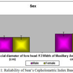 |
Figure 1: Reliability of Sear’s Cepholometric Index Based on Sex
|
Table 2: Correlation between sear’s cepholometric index and width of maxillary anterior teeth.
Correlations
| Age | Circumferencial diameter of fore head /13 (cm) | Width of Maxillary Anteriorsintraorally in cm | ||
| Age | Pearson Correlation | 1 | 0.135 | 0.212 |
| Sig. (2-tailed) | 0.572 | 0.37 | ||
| N | 20 | 20 | 20 | |
| Circumferencial diameter of fore head /13 (cm) | Pearson Correlation | 0.135 | 1 | .642** |
| Sig. (2-tailed) | 0.572 | 0.002 | ||
| N | 20 | 20 | 20 | |
| Width of Maxillary Anteriorsintraorally in cm | Pearson Correlation | 0.212 | .642** | 1 |
| Sig. (2-tailed) | 0.37 | 0.002 | ||
| N | 20 | 20 | 20 |
**. Correlation is significant at the 0.01 level (2-tailed).
Discussion
The development of a pleasing orofacial expressions of a patient depends upon the ability of the dentist to replace the denture in such a way that it replicates the natural tooth present in the arch.[9] Furnas stated that, aesthetics in full denture construction as employed by at least in 90% of men in general practice is said conspicuous by its absence[10] Many techniques exist for the selection of anterior teeth but this study aims to determine the reliability of Sear’s cepholometric index in determination of width of maxillary anterior teeth for anterior teeth selection. The present study reveals that the mean width of maxillary anterior teeth measured from left canine eminence to right canine eminence was found to be 4.9495 cm in males and 4.978 cm in females which was almost equivalent. Moreover, the mean value of circumferential diameter of forehead measured in female patient was 4.0136 cm and in male patient it was 4.0133 cm which was found to be similar. These findings indicate that there is no significant difference in mean values on gender comparison. So, Sear’scepholometric index is applicable universally on both male and female patients.This study also reveals demonstrable correlation between Sear’s cepholometric index and width of maxillary anterior teeth which is helpful in anterior teeth selection for completely edentulous patients in the absence of patients with pre-extraction records.Several other studies have been done which involves using bizygomatic width of the face as an index for maxillary anterior teeth selection. The present study has much relevance when compared to the studies done on Berry’s method[11], Harmony method[12], Nelson method[13] and Tabular Dimension Table Method[14,15] since none of the above mentioned studies uses sex as a a parameter for comparison in anterior teeth selection.
Conclusion
Sear’s cephalometric index is a highly valid indicator of anterior teeth selection. Therefore ,Sear’scephalometric index is used to assist clinicians to calculate maxillary teeth width in completely edentulous patients with absence of pre-extraction records. But other factors like colour and contour of the artificial teeth, musculature of oral mucosa, contour of residual ridge must also be considered for the success of the denture.
Aknowledgement
Authors greatly acknowledge and thank all the participants for their participation.
Conflict of Intrest
Authors states there is no conflict of intrest.
References
- Hasanreisoglu U, et al. An analysis of maxillary anterior teeth: facial and dental proportions.J Prosthet Dent. 2005;94:530–538. doi: 10.1016/j.prosdent.2005.10.007.
CrossRef - Wright WH. Selection and arrangement of artificial teeth for complete prosthetic dentures.J Am Dent Assoc. 1936;23:2291–2307.
- Land LS. Anterior tooth selection and guidelines complete denture esthetics. In: Winkler S, editor.Essentials of complete denture prosthodontics. St. Louis: Ishiyaku Euro America Inc.; 1996. pp. 200–216
- Furnas IL. Esthetics in full denture construction.J Am Dent Assoc. 1936;23:3.
- Alexander LM. Clinical applications of concepts of functional anatomy and speech science to complete denture Prosthodontics. J prosthet dent. 1963;13:204–227. doi: 10.1016/0022-3913(63)90165-3.
CrossRef - Zarb GA, Bolender CL, Hickey JC, Carlsson GE. Textbook on bouchersprosthodontic treatment for the elderly. 10. New Delhi: BI Publications Pvt Ltd; 1998. Selecting artificial teeth for the edentulous patient; pp. 330–351.
- Hoffman W, Jr, Bomberg TJ, Hatch RA. Interalar width as guide in denture tooth selection. J Prosthet Dent. 1986;55:219–221. doi: 10.1016/0022-3913(86)90348-3.
CrossRef - Gomes VL, et al. Interalar distance to estimate the combined width of the six maxillary anterior teeth in oral rehabilitation treatment. J Esthet Restor Dent. 2009;21:26–36. doi: 10.1111/j.1708-8240.2008.00227.x.
CrossRef - Wehner PJ, Hickey JC, Boucher CO. Selection of artificial teeth. J prosthet dent. 1967;18:222–232. doi: 10.1016/0022-3913(67)90185-0.
CrossRef - Kern BE. Anthropometric parameters of tooth selection. J Prosthet Dent. 1967;17(5):431–437. doi: 10.1016/0022-3913(67)90140-0.
CrossRef - Mavroskoufis F, Ritchie GM. The face form as a guide for the selection of maxillary central incisor. J Prosthet Dent. 1980;43(5):501–505. doi: 10.1016/0022-3913(80)90319-4.
CrossRef - Masuoka N, Muramatsu A, Aaiji Y, et al. Discriminative thresholds of cephalometric indexes in the subjective evaluation of facial asymmetry. Am J OrthodDentofacialOrthop. 2007;131:609–613 doi:10.1016/j.ajodo.2005.07.020.
CrossRef - Krajicek DD. Natural appearance for the individual denture patient.J Prosthet Dent. 1960;10:205. doi: 10.1016/0022-3913(60)90041-X.
CrossRef - Furnas IL. Esthetics in full denture construction. J Am Dent Assoc. 1936;23:3.

