
Manuscript accepted on :May 30, 2017
Published online on: --
Plagiarism Check: Yes
Elham Farokh-Gisour1, pegah kiani2, masoud parirokh3 and masoumeh aminizadeh4
1Department of pediatric dentistry, faculty of dentistry, Kerman university of medical sciences, Member of Department of Endodontics, Kerman University of Medical Sciences, Kerman, Iran.
2Department of Endodontics, Kerman University of Medical Sciences, Kerman, Iran.
3Department of Endodontics, faculty of dentistry, Kerman university of medical sciences, Member of Kerman Oral and Dental Diseases Research Center, Kerman, Iran.
4Department of pediatric dentistry, faculty of dentistry, Kerman university of medical sciences, Member of Department of Endodontics, Kerman University of Medical Sciences, Kerman, Iran.
DOI : https://dx.doi.org/10.13005/bpj/1186
Abstract
The aim of this study was to evaluate the presence or absence and difference of cotton size in gnashing strength of teeth filled with temporarily dressing material «coltosol». In this study, 60 human premolar teeth that had been extracted for orthodontic reasons or periodontal problems are done. Samples by stereomicroscope to leave a review before work were examined. On the teeth, access cavities for (MOD) created and for the removal of dentin tissue, teeth canals were prepared up to number 30 files. A picture was taken teeth before and after access, cavity preparation and teeth were divided randomly into four groups of 15. The first group without dressing as a control group, the second group has a cotton ball with a bandage covering only orifices (Coltosol) and the third group with a cotton ball with a bandage covering larger pulp chamber and the fourth group without a cotton ball and was just dressing. All samples were stored in room temperature water. The teeth 2500 cycle in the thermocycled were placed and then press each tooth with a Testometric and the failure threshold were recorded and analyzed by ANOVA test was used. The results showed that application of cotton under dressing lead to reduce the strength gauze is compared to lack of cotton (ANOVA, p <0.05).The results showed that application of cotton under dressing led to a reduction in the strength of the dressing material in premolars teeth. Also, by increasing the thickness of cotton, dressing strength reduced.
Keywords
Pharmacological Study; Dentistry Clinics; temporary restoration; Coltosol; Cotton ball; Compressive strength
Download this article as:| Copy the following to cite this article: Farokh-Gisour E, Kiani P, Parirokh M. Aminizadeh M. A Study Investigating the Effective Pharmacological Factors in the Strength of Filled Teeth, With A Medicinal Material. Biomed Pharmacol J 2017;10(2). |
| Copy the following to cite this URL: Farokh-Gisour E, Kiani P, Parirokh M. Aminizadeh M. A Study Investigating the Effective Pharmacological Factors in the Strength of Filled Teeth, With A Medicinal Material. Biomed Pharmacol J 2017;10(2). Available from: http://biomedpharmajournal.org/?p=15212 |
Introduction
When for any reason the treatment is considered the root of two or more visits, coronal coverage canals and crowns temporary restoration is considered among the most important challenges [1].
Diverse products as temporary restorative materials between appointments, presented root now is one of the most common materials used as dressing tooth between appointments coltosol is placed. Coltosol benefits can be used to transfer and easy, convenient and affordable coronal seal mentioned. In addition, after exposure to the oral environment, the expansion due to moisture absorption occurs in the material that makes proper compliance with the cavity walls. Coltosol disadvantages can be too much expansion can cause cracks and breakage during filling occlusal tooth, relative soft to the material that causes considerable wear it, and not having the necessary strength in large cavities, especially cavities named Class Two [2 and 3]. For various reasons, main ingredient under a temporary repair or gauze cotton balls used. Among the advantages of cotton, can be prevented from entering the dressing material into the root canal, reduce the impact on the interplay dressing ingredients and ease of root canal and the surrounding dressing material removal can be mentioned in the next session [4].
Using cotton, lower volumes of gauze material used and therefore relatively less volume changes. On the other hand, many dentists believe that cotton can be a suitable means for maintaining the desired amounts of the drug with the release of the cotton and entering the root canal during the days between sessions, dental treatment, through the effects of antimicrobial, analgesic, or so [5]. Previous studies conducted mainly on coronal microleakage following the use of different materials used for dressing teeth [1, 6] or compressive strength and failure factors after restoration of teeth (especially restoration) permanent teeth that have root canal treatment [3, 7, 8] are focused. However, such questions as whether the size of cotton gauze is placed below the compressive strength of teeth and whether size cotton too Coltosol affect the expansion so far unanswered.
In recent years, little work in the field of cotton under the dressing has been done. Krakow et al. (1977) and Lamers et al (1980) clinical studies conducted on the benefits of cotton under the dressing. In this regard, Messer & Wilson (1996) have proposed a method occlusal cavity. They concluded that while the dressing material, pulp chamber and cavity walls should be dry [3]. In another clinical study, Sjogren et al (1991) of a sterile foam layer temporary restoration for more than five weeks, but without flood dressing compromise. However, they indicated that the use of a cotton layer could have side effects that are at risk to flooding. 1) Cotton temporary restoration substantially reduces and the leakage is increased. 2) Cotton by acting as a pillow, which allows movement during chewing movements, gives strength to endanger dressing 3) cotton gauze material distance between the wall and throws. 4) Cotton fiber pellets may stick to the walls of the cavity. 5) Cotton increases the risk of leakage between the exposed side channels [9]
Naseri et al (2012), three dressing: Coltosol, Cavizol and Zonalin of the coronal seal during different times compared. The result of the study implies that all materials showed leakage increases over time.
However, Coltosol and Cavizol more suitable material to create a coronal seal in the intervals are longer than a week. [8] Charles et al (2002) the importance of using cotton ball looked after canal filling. For this purpose, 46 endodontists and 46 general dentists chosen, 40 Endodontics (87%) and 25 general practitioners (54%) to the question “Is there a general dentist cotton ball in the pulp chamber after filling channel needs” responded. Endodontists 25 of 40 patients (62.5%) and 20 general practitioners from 25 patients (80%) said they think a general dentist needs a cotton ball in the pulp chamber. The proportions of these two groups showed no significant difference according to Chi-square test. [10] The aim of this study was to evaluate the effect of cotton on the strength of its effect on morphology of the teeth gnashing of teeth is observed. In the study of factors associated with tooth crown fractures treated, the extent of the material, remain intact marginal ridge of the mesial and distal, a person’s age, type (number) of teeth, fillings previous, the need to Endo, the remaining walls, among the factors listed and cervical wear [12, 5, 11] and therefore in the design of this study considered.
Materials and Methods
This study, as a study of experimental-blind type, 60 human premolar teeth was used for this purpose. Dental health are examined under stereomicroscope with magnification 20 and were evaluated using the catheter tip and in case of any breakage, cracks, corrosion, wear, remaining pressure of forceps, or any anomaly in the crown were excluded. Teeth that were perfectly healthy and did not have these problems again were kept in containers of 20% formalin solution. Periodontal gum tissue or teeth that have remained attached to their surfaces, by scaling pison, at least they were clean power. Collect teeth, as described above until the number of 70 teeth with ideal conditions continued and achieved. Access cavity preparation teeth before photography from four angles buccal, lingual, mesial and distal prepared. Selected teeth root canal access cavities were prepared by turbines. Fisher diamond drills holes with a diameter of 1 and 8 mm high speed turbines were prepared. After checking milled, the soft tissue inside large pulp chamber by the handpiece with a very slow round and soft tissue coronal half of the root canal of each tooth using a hand file to number 30 and rinse with tap water were removed. Access cavity preparation and cleanup process continues until favorable conditions achieved with 60 teeth then the teeth were taken for the correctness of this issue with the other. The 60 randomly into four groups of 15 rats using the draw were divided as follows:
Group A: teeth with cavities access without any changes
Group B: teeth with cotton pellet holes + access so that only covered orifices + dressing material
Group C: teeth with access holes to the size of the pulp chamber cover + cotton + gauze material
Group D: teeth with cavities access + the dressing that covers all pulp chamber access holes.
After teeth are divided into four groups to do the next steps on them, to ensure homogeneity of the four measurements were performed on it as follows:
The thickness of the buccal and lingual walls teeth using a dial caliper with a precision of 0.1 mm evaluated from different parts and the minimum thickness was recorded that occur in each wall. The average thickness of the walls of the buccal and lingual teeth four groups were compared using independent t-test and significant difference was observed between the groups. the remaining wall thickness mesial and distal pulp chamber of enamel and cementum junction (CEJ) using a calibrated caliper with a precision of 0.1 record and the average numbers recorded were compared using independent t-test, in which case the difference was not significant.
Teeth Group A without performing any other steps, until the evaluation of fracture force, were kept in normal saline. In the teeth of Group B was initially a small cotton ball and then the teeth with Article Colotosol (manufactured by Aria Dent, Tehran, Iran) were filled. In the teeth of Group C of 8 small cotton ball (instead of one Group B) were used.
Use several small cotton ball instead of a big shot, due to the random selection of them) would ensure uniformity approximate size of the group and better dental cotton used in the cover of the hole.
After eight rounds of the cotton, the material Colotosol for temporary restoration of teeth, in a similar way to what was described for Group B were used. Group D pulp chamber of teeth in all holes and access holes were filled with material Colotosol. The external surface in a way that was explained for Group B was performed. All teeth, to reconstruct the actual conditions of the oral cavity in the lab, inside containers of distilled water mixed with antifreeze and 2,500 rounds, each round of 90 seconds duration each for 40 seconds. And the distance between each bath was 10 seconds the device was thermocycled and a temperature of 5 to 60 ° rotation. The samples were removed for Testometric machine was under pressure. Pressures were recorded in the first tooth breaking point. For data analysis SpSS software version 19 was used.
Results
With respect to operations and calculations on four groups A, B, C and D descriptive statistics related to the pressure shown in Table A and Figure 1.
Table 1: descriptive statistics related to the pressure
| Standard error (N mm-2) | Standard deviation
(N mm-2) |
Average
(N mm-2) |
Maximum
(N mm-2) |
Minimum
(N mm-2) |
Number | Group |
| 40.49 | 156.84 | 376.81 | 686.70 | 175.50 | 15 | D |
| 20.00 | 77.48 | 202.09 | 401.90 | 115.20 | 15 | C |
| 29.28 | 113.40 | 270.19 | 565.80 | 144.20 | 15 | B |
| 19.61 | 75.97 | 152.68 | 281.10 | 66.50 | 15 | A |
| 17.73 | 137.35 | 250.44 | 686.70 | 66.50 | 60 | Total |
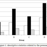 |
Figure 1: descriptive statistics related to the pressure
|
Comparison of four groups according to ANOVA test showed a significant difference between the pressure in the four companies
F value = 11.477 and (p-value = .000) after Bonferroni test indicated a significant difference between group 1 with two and four (p <0.05).
There was also a significant difference between the groups of three and four (p <0.05). Table 2
Table 2: significant difference between the groups of three and four
| (I) group | (J) group | p-value. |
| D | 2 | 0 |
| 3 | 0.066 | |
| 4 | 0 | |
| C | 1 | 0 |
| 3 | 0.59 | |
| 4 | 1 | |
| B | 1 | 0.066 |
| 2 | 0.59 | |
| 4 | 0.032 | |
| A | 1 | 0 |
| 2 | 1 | |
| 3 | 0.032 |
According to the statistical results obtained and the average of the four Group D, C B, A Myagyn pressure in Group A is minimal and significant difference with other groups and Group C with four significant differences in the mean pressure is obtained.
Discussion and Conclusion
This study aimed to evaluate the strength of Coltosol dressing following application of the dressings was made of cotton with different sizes. The results showed that the addition of cotton in the dressing leads to a reduction in the strength of temporary restoration and increasing the thickness of the dressing cotton, as well as strength greater reduction will be temporary restoration.
In relation to the use of temporary repair and restoration of root canal treatment in between sessions, few studies have been done. Also, studies regarding the application of the dressing cotton are also very low, the present study was conducted to evaluate the effect of cotton under temporary restoration Coltosol. In a study by Charles et al (2002) performed, comments endodontists and general dentists regarding the use of cotton in the tooth after root canal filling was examined. The results of their study showed that more than Endodontists are dentists believe that this issue should be a general dentist after root canal treatment, dental pulp with cotton filled [10]
Bruce et al (2001) used cotton leakage under the temporary restoration examined. They observed that thick cotton leads to increased leakage of the temporary restoration and the study findings can be linked so that the increase in the thickness of cotton led to a reduction in the strength of the present study was dressing. As mentioned earlier, the use of cotton in the dressing can be significantly resulting in increased leakage and this clinical decisions regarding the application of the dressing makes it harder cotton [12]. Lausten et al (2005) showed that the use of dressing Coltosol due to expansion caused by the setting temporary restorative material, compared with ZOE led to an increase in Kaspi broken and cracked teeth by turning the MOD. This is also the long-term Coltosol faced with the question; in addition to the application of the dressing cotton was also questionable [5].
Among the limitations of this study, failure to investigate the microleakage following the use of thin cotton or gauze is much below. It is recommended that future studies, leakage of cotton in the dressing following application will also be assessed. Overall results showed that application of the dressing cotton led to a reduction in the strength of the dressing material in premolars teeth. Also, by increasing the thickness of cotton, dressing, reduced strength. It is recommended that the following studies related to the leakage cotton dressing following application will also be made to cotton of applying the dressing is quite reasonable to reject or approve.
Refrences
- Dillard CR, Barfield RD, Tilashalski KR, Chavers LS, Eleazer PD. Comparison of endodontist versus generalist regarding preference for postendodontic use of cotton pellets in pulp chamber. J Endod. 2002;28(9):656-7.
CrossRef - Endodontists AAo. Coronal Leakage: Clinical and Biological Implications in Endodontic Success. 2002.
- Lamers AC, Simon M, van Mullem PJ (1980) Microleakage of Cavit temporary flling material in endodontic access cavities in monkey teeth. Oral Surgery, Oral Medicine, Oral Pathology 49,541^3.
CrossRef - Gomes-Filho JE, Aurélio KG, Costa MM, Bernabé PF. Comparison of the biocompatibility of different root canal irrigants. J Appl Oral Sci. 2008;16(2):137-44.
CrossRef - Laustsen MH, Munksgaard EC, Reit C, Bjørndal L. A temporary filling material may cause cusp deflection, infractions and fractures in endodontically treated teeth. Int Endod J. 2005;38(9):653-7.
CrossRef - KrakowAA, destoppelaar JD, GrUn P (1977)In vivo study of temporary ¢lling materials used in endodontics in anterior teeth.Oral Surgery, Oral Medicine, Oral Pathology43,615^20
CrossRef - Madarati A, Rekab MS, Watts DC, Qualtrough A. Time-dependence of coronal seal of temporary materials used in endodontics. Aust Endod J. 2008;34(3):89-93.
CrossRef - Naseri M, Ahangari Z, Shahbazi Moghadam M, Mohammadian M. Coronal sealing ability of three temporary filling materials. Iran Endod J. 2012;7(1):20-4.
- Orahood JP, Cochran MA, Swartz M, Newton CW (1986)In vitro Study of marginal leakage between temporary sealing materials and recently placed restorative materials .Journal of Endodontics12,523^7.
- Charles R. Dillard, Robert D. Barfield, Ken R. Tilashalski, L. Scott Chavers, and Paul D. Eleazer(2002) Comparison of Endodontist Versus Generalist Regarding Preference for Postendodontic Use of Cotton Pellets in Pulp Chamber. JOURNAL OF NDODONTICS. VOL. 28, NO.9.
- Messer HH, Wilson PR (1996) Preparation for restoration andtemporization. In :Principles and Practice of Endodontics,2nd edn. Philadelphia, USA:W.B. Saunders Co., pp. 260^76.
- Newcomb BE, Clark SJ, Eleazer PD. Degradation of the sealing properties of a zinc oxide-calcium sulfate based temporary filling material by entrapped cotton fibers. J Endodon 2001;27:789 –90.
CrossRef

