
Manuscript accepted on :March 10, 2017
Published online on: --
Plagiarism Check: Yes
Satheesh Babu1, Sudhakar1, E. Rajesh2 and N. Anitha2
1Department of Oral Pathology, Sree Venkateswaraa Dental College and Hospital, Ariyur. Puducherry.
2Department of Oral Pathology, Sree Balaji Dental College and Hospital, Bharath University, Pallikaranai, Chennai–600100.
DOI : https://dx.doi.org/10.13005/bpj/1126
Abstract
Lateral periodontal cyst (LPC) is an uncommon type of developmental odontogenic cyst that typically occurs laterally on the root surface of a tooth, representing 0.8% of cysts in the jaws. The objective of this article was to describe the diagnosis of LPC based on the clinical, radiographic examination, and histopathologicanalysis, and to display the treatment of the case. The clinical, radiographic, and histopathological examinations were carried out in the case confirming the diagnosis of LPC. Surgical enucleation was performed on the lesion. In the follow-up of the case there was complete bone regeneration and no recurrences. Although the occurrence of lateral periodontal cyst is rare, the precision of its diagnosis is necessary so that the correct treatment can be established.
Keywords
Lateral periodontal cyst; Odontogenic Cyst
Download this article as:| Copy the following to cite this article: Babu S, Sudhakar S, Rajesh E, Anitha N. Lateral Periodontal Cyst _ A Case Report. Biomed Pharmacol J 2017;10(1). |
| Copy the following to cite this URL: Babu S, Sudhakar S, Rajesh E, Anitha N. Lateral Periodontal Cyst _ A Case Report. Biomed Pharmacol J 2017;10(1). Available from: http://biomedpharmajournal.org/?p=14126 |
Introduction
The lateral periodontal lateral cyst is a developmental odontogenic cyst defined as a radiolucent lesion, along the lateral aspect of an erupted vital tooth in which an inflammatory etiology and a diagnosis of collateral keratocyst have been excluded based on clinical and histological grounds (1).
Since the last maxillary cyst classification of the World Health Organization (WHO), described by Kramer et al. in 1992, lateral periodontal cysts (LPCs) have been regarded as an independent condition (2).
The clinical manifestations tend to be mild, and the diagnosis is generally established by means of a routine radiological exploration, which reveals a radiolucent image with less than 1 cm in size in most cases (3-9). LPCs account for between 0.8% (3) and 1.5% (4) of all maxillary cysts.
Case Discussion
A 20 year female patient came with a complaint of swelling in the upper front tooth region for the past 2 years. On intra oral examination a swelling was present in the labial aspect extending from 11 to 13 region (fig 1) which was asymptomatic.
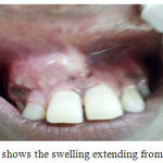 |
Figure 1: shows the swelling extending from 11 to 13. |
Panoramic examination showed a well defined radiolucency which was present in between the middle and apical third of 12 and 13 with divergence of roots ( fig 2 and 3).
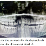 |
Figure 2: Showing panoramic view showing a unilocular radiolucency with divergence of 12 and 13. |
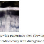 |
Figure 3: showing panoramic view showing a unilocular radiolucency with divergence of 12 and 13. |
The swelling was aspirated which contained a yellowish fluid (fig4). The tissue was measuring 1.5 cm * 1cm Surgical enucleation of the cyst was done and sent for histopathologic evaluation . (fig4).
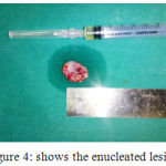 |
Figure 4: shows the enucleated lesion. |
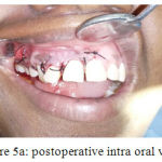 |
Figure 5a: postoperative intra oral view |
The H and E stained soft tissue section showed a stratified squamous lining epithelium consisting of 8 – 10 cell layer thickness (fig 5 and 6). The epithelial connective tissue interface was flat and devoid of rete ridges. The epithelium showed areas of spongiosis. The stroma was loosely arranged with minimal inflammation.
The histopathologic evaluation was done to rule out any malignancies.
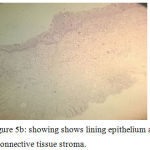 |
Figure 5b: showing shows lining epithelium and a connective tissue stroma. |
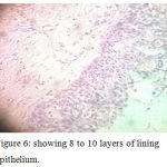 |
Figure 6: showing 8 to 10 layers of lining epithelium. |
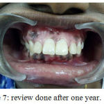 |
Figure 7: review done after one year. |
Discussion
The lateral periodontal cyst is a developmental odontogenic cyst which is usually uncommon. It is located along the lateral aspect of a root of a vital tooth. In this case it was seen in the lateral aspect of 12 and 13 region. Lesions has been removed by surgical enucleation. Special care should be taken not do damage the roots of the adjacent teeth. Recurrence is uncommon but has sporadicallly been reported (12). Here the patient was reviewed periodically ( fig7) and no evidence of recurrence.
References
- Text book of cysts – Shear.
- Kramer IRH, Pindborg JJ, Shear M. WHO Histological Typing of Odontogenic Tumours. 2nd ed. Geneva: Springer-Verlag; 1992. p.34-118.
- Altini M, Shear M. The lateral periodontal cyst: an update. J Oral Pathol Med. 1992 Jul;21(6):245-50.
- Kerezoudis NP, Donta-Bakoyianni C, Siskos G. The lateral periodontal cyst: aetiology, clinical significance and diagnosis. Endod Dent Traumatol. 2000 Aug;16(4):144-50.
- Angelopoulou E, Angelopoulos AP. Lateral periodontal cyst. Review of the literature and report of a case. J Periodontol. 1990 Feb;61(2):126-31.
- Carter LC, Carney YL, Perez-Pudlewski D. Lateral periodontal cyst. Multifactorial analysis of a previously unreported series. Oral Surg Oral Med Oral Pathol Oral Radiol Endod. 1996 Feb;81(2):210-6.
- Lindh C, Larsson A. Unusual jaw-bone cysts. J Oral Maxillofac Surg. 1990 Mar;48(3):258-63.
- Rasmusson LG, Magnusson BC, Borrman H. The lateral periodontal cyst. A histopathological and radiographic study of 32 cases. Br J Oral Maxillofac Surg. 1991 Feb;29(1):54-7.
- Tolson GE, Czuszak CA, Billman MA, Lewis DM. Report of a lateral periodontal cyst and gingival cyst occurring in the same patient. J Periodontol. 1996 May;67(5):541-4.
- Lateral periodontal cyst A case report and review of literature. Luis feliepe das . Journal of oral and maxillofacial research.
- Two cases of lateral periodontal cyst . oral surgery oral medicine and oral pathology feb 1967 vol 2392) pg 183-188.
- Greer RO, Johnson M. Botryoid odontogenic cyst: clinical pathologic analysis of teh cases with three recurrences. J Oral Maxillofac Surg 1988;46:574-579.

