
Manuscript accepted on :March 03, 2017
Published online on: --
Plagiarism Check: Yes
Raza Mohamed1, Noufal P. K2, Deepa Shenoy3, P. Sesha Reddy4, ACU. Varma4 and Ashish R. Jain5
1Department of Prosthodontics, Pariyaram dental college, Pariyaram, Kannur(Dt), kerala, India.
2Department of Prosthodontics, Al Azhar Dental College, Perumpillichira, Thodupuzha, Kerela, India.
3Department of Prosthodontics, Asan Memorial Dental College and Hospitals,Chennai, India.
4Department of Prosthodontics, Goverment Dental College, RIMS, Putlampali, Kadapa, India.
5Department of Prosthodontics, Saveetha Dental College and Hospitals,Chennai, India.
Corresponding Author E-mail: dr.ashishjain_r@yahoo.com
DOI : https://dx.doi.org/10.13005/bpj/1128
Abstract
The fractures and debonding of acrylic resin teeth from acrylic denture base is a common clinical problem. This bond may be further enhanced by removing the glossy ridge lap surfaces of these teeth, or by providing mechanical retention grooves.The purpose of this study was to compare the tensile bond strength of a conventional denture base resin to a cross-linked acrylic teeth processed under two different curing cycles, having three variables in each group. Fourty eight specimens were prepared for tensile bond strength testing using Instron Universal testing machine under two different curing cycles. Group I: Glossy surface of the ridge-lap area of the acrylic teeth unmodified, Group II: Glossy surface of the ridge-lap area of the acrylic teeth abraded, Group III: Preparation of retentive grooves on the lap area of the acrylic teeth.The tensile bond strength of specimens cured using slow curing cycle gave a better result when compared to the short Curing cycle and the slow curing cycle with retentive grooves was found to have the highest tensile bond strength among the other variables.Overall, higher bond strength of denture base resin to the denture teeth tested were obtained with the slow curing cycle rather than short curing cycle. The bond between the acrylic teeth and denture base was improved significantly by the preparation of retentive grooves on the ridge lap area compared to the ridge lap area abraded and when left untreated. The strongest bond was with the specimens processed under the slow curing cycle with retentive grooves placed on the ridge lap area of the tooth.
Keywords
Artificial Teeth; Bond Strength; Curing; Denture Base
Download this article as:| Copy the following to cite this article: Mohamed R, Noufal P. K, Shenoy D, Reddy P. S, Jain A. R, Varma A. A Comparitive Study on the Tensile Bond Strength of Conventional Denture Base Resin to Cross Linked Acrylic Tooth using two Different Curing Cycles - an invitro Study. Biomed Pharmacol J 2017;10(1). |
| Copy the following to cite this URL: Mohamed R, Noufal P. K, Shenoy D, Reddy P. S, Jain A. R, Varma A. A Comparitive Study on the Tensile Bond Strength of Conventional Denture Base Resin to Cross Linked Acrylic Tooth using two Different Curing Cycles - an invitro Study. Biomed Pharmacol J 2017;10(1). Available from: http://biomedpharmajournal.org/?p=13758 |
Introduction
The original acrylic denture teeth introduced in 1940 were esthetic and easy to adjust but were susceptible to crazing. Denture base materials were made from acrylic resin as early as 1937 1. These denture bases could craze and fracture easily. Cross-linking helped to sovle the problem of crazing, but made bonding to acrylic teeth more difficult. Several factors affect the bond including cross-linking of materials, contamination during processing, and available monomer during processing. Poly (methyl methacrylate) is the most frequently used denture base material to the present date. Soon after its introduction, acrylic resin became popular as denture base material that within ten years, 98% of all the dentures were constructed of methyl methacrylate polymers. Since then other types of polymer or co polymer have been developed to be used as denture base materials. As a known fact, the polymerization processes useful for dental resins are commonly activated by one of the following methods: heat, chemical, light and microwave energy.2-5 Of these methods heat curing has one great advantage over the others is that, an increased rate of monomer diffusion occurs at higher temperature which leads to better wetting of cross linked acrylic teeth by the dough. There are two alternative heat-curing techniques based on the rate at which curing occurs: slow curing and short curing. Slow curing result in much tougher denture bases, producing fewer cross links and branches, and having a higher overall molecular weight between cross links because fewer polymer chains grew at any time. However, the main disadvantage of this curing cycle is the time factor involved. Short curing cycle, which involves too rapid rise in temperature, produces large number of radicals resulting in many growing polymer chains. This leads to the building up of heat in the dough until the boilding point of the monomer is exceeded, which results in porosity with subsequent loss of strength, toughness and esthetics 6-9. The fractures and debonding of acrylic resin teeth from acrylic denture base is a common clinical problem. As many as one third of denture repairs were due to debonding of teeth from denture base 11-12. The degree of cross-linking within the acrylic teeth is greater than that within polymerized denture bases. A chemical bond is achieved between the acrylic teeth and the denture base resin because of the reduced cross-linking seen at the cervical portions of these teeth. This bond may be further enhanced by removing the glossy ‘ridge lap’ surfaces of these teeth, or by providing mechanical retention grooves13-18. The purpose of this study was to compare the tensile bond strength of a conventional denture base resin to a cross-linked acrylic teeth processed under two different curing cycles, having three variables in each group.
Materials and Methods
This study was conducted to evaluate the comparative tensile bond strengths of conventional heat cured denture base resin to cross-linked acrylic teeth using two different curing cycles- 1) The short curing cycle and 2) the slow curing cycle. Three variables were used in each curing cycle and they were named as Group I, Group II and Group III.
Group I: Glossy surface of the ridge-lap area of the acrylic teeth unmodified.
Group II: Glossy surface of the ridge-lap area of the acrylic teeth abraded.
Group III: Preparation of retentive grooves on the lap area of the acrylic teeth.
Preparation of the Teeth
Only one brand [upper right central incisor of premadent co.32 mold] of cross linked acrylic teeth were used in this study. In order to standardize the tooth size, only the same molds of anterior teeth were chosen for this study. In group I, the glossy surfaces of the ridge lap areas were left unmodified. These were used as the control groups in both the curing cycles. In Group II, the glossy surfaces of the ridge lap areas of the teeth were abraded. The surface was abraded to an extent where the gloss would disappear, using a 160 no. Sandpaper in one direction with the same frequency. In Group III, retentive grooves were placed on the ridge lap surface of the teeth. Retentive grooves of 2mm depth and 2mm width were cut along the entire mesio-distal length of the tooth, using an inverted cone bur. A steel split mould (Fig.1 and 2) was used to obtain a wax pattern in the shape of a rod of uniform diameter of 7mm and 35mm length. These rods were used so that the specimens were uniform in size and for the case of fabrication of the moulds. The acrylic teeth from all the three groups were attached to one end of the wax rod and wax pattern were made in such a way that only the ridge lap area of the teeth were in contact with wax (Fig. 3 and 4). The teeth were placed straight, without giving any inclination. The excess wax was carved to simulate a clinical condition (the position in which a tooth is placed on a waxed up trial denture base). The resultant wax patterns and attached teeth were invested in type III dental stone, dewaxed, and packed with heat cure denture base material (DPI, type I) and flasked in a standard dental flask in a manner similar to that used in the fabrication of a conventional acrylic denture. Latex examination gloves were used to avoid contamination of the mixed resin during packing. Care was taken to ensure that the tooth position was not disturbed during the entire process. The flasks were bench cured for one hour after which they were cured, one according to the slow curing cycle.
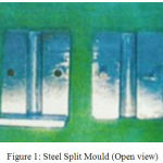 |
Figure 1: Steel Split Mould (Open view)
|
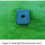 |
Figure 2: Steel Split Mould (closed view)
|
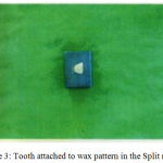 |
Figure 3: Tooth attached to wax pattern in the Split mould
|
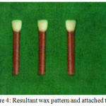 |
Figure 4: Resultant wax pattern and attached tooth
|
Short Curing Cycle
In this curing cycle, the flask was immersed in water at room temperature and then the temperature was raised to 75 degrees centigrade (167.2F) and was allowed to polymerize for 90 minutes. The temperature was then raised to 100 degree centigrade and maintained for 60 minutes.
Slow Curing Cycle
In this curing cycle, the flask was immersed in water at room temperature and the temperature was raised to 75 degrees centigrade (167.2F) and processed for 8 hrs.
After curing, the flasks were allowed to bench cool overnight. The flasks were then opened and the cured specimens were retrieved. The retrieved specimens were finished with a fine stone. The specimens were then stored in water for 24 hrs. So that the residual monomer present on the surface of the cured specimens could leach out. (fig 5)
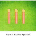 |
Figure 5: Acrylized Specimen
|
Tensile Bond Strength Testing of the Specimens
Tensile bond testing was performed on an Instron Universal testing machine model 1125 [Instron limited]. Instron Grips were used to hold the specimen firmly during testing (Fig.6). The specimens were inserted into the grips with the upper and lower members tightened equally and tension was applied axially with a cross. Head speed of 0.05cm/min until failure occurred (Fig. 7 and 8) and failure load was measured for each specimen
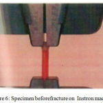 |
Figure 6: Specimen beforefracture on Instron machine
|
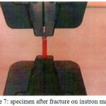 |
Figure 7: specimen after fracture on instron machine
|
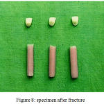 |
Figure 8: specimen after fracture
|
Fourty eight specimens were prepared for tensile bond strength testing. Out of which twenty-four were processed through short heat curing cycle and the remaing twenty-four were processed through slow heat curing cycle. There were three variables in each cycle. Each variable had eight specimens to be tested. The results of bond stength thus obtained were measured in kilograms for each specimen and recorded as the total load. They were converted to kg/cm2 as follows.
Load =force in kg/cm2
Where area =nr2 (r is the radius of the specimen).
All the readings were recorded in kg/cm2 and were tabulated in tables I to IX.
Data was analyzed using the statistical package SPSS/PC. One-way analysis of variance followed by the multiple range tests was used for comparing the tensile bond strengths of different groups. A P value<0.05 was considered as significant.
Results
The results of bond stength thus obtained were measured in kilograms for each specimen and recorded as the total load. The maximum bond strength recorded in Group I was 99.25 kg/cm2 and the minimum bond strength recorded was 80.69 kg/cm2. Group II recorded maximum bond strength of 168.76 kg/cm2 and minimum of 154.29 kg/cm2. Group III maximum of 179.56 kg/cm2 and a minimum of 159.75 kg/cm2. (Table 1). The maximum bond strength recorded in Group I was 140.25 kg/cm2 and the minimum was 128.46 kg/cm2. Group II recorded a maximum of 180.42 kg/cm2 and a minimum of 159.83 kg/cm2 Group III recorded a maximum of 194.75 kg/cm2 and a minimum of 183.42 kg/cm2 (Table 2). Group I processed under slow curing cycle had mean tensile bond strength of 136.93 kg/cm2 and a standard deviation of 3.713 the P value was found to be p<.001, statistically singificant. Group II processed under short curing cycle had mean tensile bond strength of 159.60 kg/cm2 and a standard deviation of 173.36 kg/cm2 and a standard deviation of 9.070, P value was found to be p<.oo1, statistically significant. Group III processed under short curing cycle recorded mean tensile bond strength of 167.71 kg/cm2 and standard deviation of 6.400. Group III proceed under slow curing cycle showed mean bond strength of 190.42 kg/cm2 and a standard deviation of 3.854. The P value recorded was <.001 and was found to be statistically significant. (Table 3). The mean tensile bond strength in group I was 88.999 kg/cm2 and standard deviation of 5.084. Group III had mean tensile bond strength of 167.712 kg/cm2 and standard deviation of 6.400, the P value was <.001, statistically significant (Table III). The mean tensile bond strength recorded for Group I was 136.938 kg/cm2 and a standard deviation of 3.713. Group III had mean tensile bond strength of 190.422 kg/cm2 and standard deviation of 3.854, the P value was <.001 statistically significant (Table III). The tensile bond strength of specimens cured using slow curing cycle gave a better result when compared to the short Curing cycle and the slow curing cycle with retentive grooves was found to have the highest tensile bond strength among the other variables.
Discussion
The tensile bond strength of the denture base material to a cross- linked acrylic tooth using the short curing and slow curing cycles were tested. The glossy surface of the ridge lap area untreated, i.e.Group I recorded a mean bond strength of 88.99 kg/cm2. In the most of the specimens, the failure occurred in between the tooth and resin interface. Most of bond failures were adhesive bond failures in the form of sepration. The glossy surface of the ridge lap area abraded i.e Group II showed an increase in the mean tensile bond strength. This suggests that a proper abrasion of the ridgelap surface of a cross-linked acrylic tooth increased the bond strength. In Group III, when retentive grooves of 2mm width and 2mm depth were placed on the entire mesio-distal length of the ridge lap area, a marked increase in the bond strength was seen when compared to the ridge-lap area untreated. It suggests that the grooves might increase the surface area of the ridge lap area, thereby increasing the bond strength 7-9. In-Group III, most of the specimen showed cochesive bond failure. This may also suggest that the bond strength in itself was good and the tooth or the denture base had reduced strength. The result must be increased with caution since cochesive failure of the material may not give the exact bond strength to the denture base but it can be interpreted as the bond strength to acrylic resin exceeding the tensile failure value 12-17. None of the heat-cured specimens crossed the minimum bond strength recommended by ADA (315 kg/cm2) to pass the ADA test. It could be attributed to the variance of the testing machine or the material itself.19-21
Table 1: Tensile bond Strengths of the Specimens Processed by Short Curing Cycle in Kg/cm2
| SHORT CURING | |||
| Specimen no | Ridge-lap unaltered group I | Ridge-lap Abraded group II | Retentive Grooves on Ridge-lap group III |
| 1 | 85.46 | 165.25 | 165.46 |
| 2 | 99.25 | 168.26 | 175.42 |
| 3 | 80.69 | 159.84 | 178.85 |
| 4 | 85.49 | 160.75 | 179.56 |
| 5 | 94.69 | 159.48 | 168.75 |
| 6 | 92.29 | 166.85 | 164.28 |
| 7 | 90.45 | 154.29 | 160.45 |
| 8 | 89.53 | 168.76 | 159.75 |
Comparison of the tensile bond strength of Group I (ridge lap area untreated) to Group III (retentive grooves being placed on the ridge lap area) processed with short curing cycle shows that Group III recorded a higher mean bond strength of 167.712 kg/cm2 compared to 88.999 kg/cm2 for Group I with a Pvalue of more than 0.001, the difference in bond strengths between the specimens in GroupI and III (short curing) were statistically significant. Slow curing results in much tougher denture bases, producing fewer cross-links and branches. Free monomer content is lower, because of the steadier rise in internal viscosity of the curing polymer allows the monomer easier access to the growing free radicals4-7. The glossy surface untreated i.e.Group I. when compared, the mean bond strength recodred in-Group I of slow curing cycle was higher than in Group I of short curing cycle (Table-3). This suggests that the tensile bond strength was improved because of the slow during cycle, P value of more than 0.001 between groups suggested that it was statistically signaficant.
Table 2: Shows Tensile bond Strengths of the Specimens Processed by Slow Curing Cycle in Kg/cm2
| SLOW CURING | |||
| Specimen no | Ridge-lap unaltered group I | Ridge-lap Abraded group II | Retentive Grooves on Ridge-lap group III |
| 1 | 128.46 | 175.46 | 183.42 |
| 2 | 140.25 | 172.31 | 190.43 |
| 3 | 136.46 | 180.42 | 191.64 |
| 4 | 137.82 | 175.43 | 192.75 |
| 5 | 134.86 | 164.45 | 190.83 |
| 6 | 139.85 | 163.45 | 194.75 |
| 7 | 132.76 | 167.45 | 188.73 |
| 8 | 133.56 | 159.83 | 184.65 |
Group III (slow curing cycle) i.e. with retentive grooves showed the highest mean tensile bond strength between both the curing cycles (Table-2). The mean tensile bond strength was greater in Group III (slow curing) then in-Group II (slow curing). A.P value of more than 0.001 was obtained between Group III, thus showing a statistically significant result. This proved that the bond strength can be markedly increased by placing grooves on the ridge lap area of the tooth. When Groups III were compared, (i.e.retentive grooves placed on ridge-lap area in the short curing and the slow curing cycles) group III (slow curing) showed a significant increase in the tensile bond strength.
Table 3: Comparison between short curing vs slow curing
| Group | Class | Mean | Std. Deviation | t | |
| Ridge lap area untreated | Short curing
Slow curing |
88.9993
136.9387 |
5.0843
3.7137 |
29.4890
P<.001 vhs |
|
| Ridge lap area abraded | Short curing
Slow curing |
159.6067
173.3600 |
5.8760
9.0704 |
4.9290
P<.001 vhs |
|
| Retentive grooves on ridge lap | Short curing
Slow curing |
167.7127
190.4220 |
6.4008
3.8540 |
11.7720
P<.001 vhs |
|
Comparing the mean tensile bond strength of both the curing cycles, it was found that the slow curing had a better bond strength. However, comparing, in between the variables, slow curing, with retentive grooves placed on the ridge lap area was found to have the maximum tensile bond strength (Table 3). As the specimens processed under short curing cycle with glossy ridge lap area exhibited lower bond strengths and most of the recorded failures were adhesive, the nature of the bond between these teeth and denture base resins may increase the probability of denture tooth displacement from the prosthosis during function11-16. The result of this study points out the need to further examine the tooth resin interface after failure by scanning electron microscope to determine the penetration of the resin into the retentive grooves of the ridge lap area.
Conclusions
Within the limitations of this study, overall, higher bond strength of denture base resin to the denture teeth tested were obtained with the slow curing cycle rather than short curing cycle. The bond between the acrylic teeth and denture base was improved significantly by the preparation of retentive grooves on the ridge lap area compared to the ridge lap area abraded and when left untreated. The strongest bond was with the specimens processed under the slow curing cycle with retentive grooves placed on the ridge lap area of the tooth
References
- Anderson J.N.; strength of the joint between the plain and copolymer acrylic teeth and denture base Resins. BrDent J 1958; 6:317 – 320.
- Anthony D.H. and peyton F.A.: Dimensional Accuracy of Various Denture Base Materials. J Prosthet Dent 1962; 12:67 – 81.
- Barpal D., Curtis D.A., Finzen F, Perry J. and Gansky S.A: failure load of acrylic resin denture teeth bonded to high impact acrylic resin. J. Prosthet. Dent. 1998; 80:666 – 671.
- Buyuyukyilmaz S., Ruyter I.E: The effects of polymerzation temperature on the acrylic resin denture base-tooth bond. Int. J. prosthodont. 1997; 10:49 – 54.
- Cardesh H.S., leberman R. And Helft M.: Effect of retention grooves in acrylic resin teeth on tooth denture-base bond. J. Prosthet dent 1986; 55: 526 – 528.
- Chung R.W., Clark R.K. and Darwall B.W.: bonding of acrylic cold cure resin to acrylic denture teeth. Aust dent J 1995; 40: 241 – 245.
- Clancy J.M.S., L.Foster Hawkins, john C.Keller and Dan B.boyer: bond strength and failure analysis of light cured denture resins bonded to denture teeth. J prosthet dent 1991; 65: 315 – 324.
- Cornell J.A., Tucker J.L. and Power C.M.: Physical Properties of Denture base materials. J Prosthet Dent 1960; 10:516 – 524.
- Darbar U.R., Huggest R., Barrison A. And williams K.: Finite element analysis of stress distribution at the tooth-denture base interface of acrylic resin teeth debonding from the denture base. J Prosthet dent 1995; 74: 591- 594.
- Dicracy F.V.: Influence of certain variables on the toothbrush abrasion of resin veneering materials. J Prosthet dent 1962; 12: 720 – 731.
- Hans T., Ejvind Budtz-Jrgesen, Odont D.R. and Ulrik Bertram: A four-year follow up study on processed pour acrylic resins. J prosthet Dent 1980; 44: 495 – 496.
- Marcheck B.W., Yu-Z; zhao-X.Y. and White-S.N.: Adhesion of denture tooth porcelain to heat polymerized denture resin. J Prosthet dent 1995; 74:242 – 249.
- Marrow R.M, Matvis F.M, Windeles A.S. and Fuchs R.J: bonding of plastic teeth to tow heat curing denture base resins J.Prosthet dent 1978;39:565-568.
- Smith D.C.: The acrylic denture base. Mechanical evaluation of midline fractures. Br Dent J 1961; 110: 257 – 267.
- Sprately M.H.: An investigation of the adhesive of acrylic resin teeth to dentures. J Prosthet dent 1987; 58: 389 – 392.
- Takahashi Y., Chai J, Takahashi T, Habu T. Bond strength of denture teeth to denture base resins. Int J Prosthodont. 2000 Jan-Feb;13(1):59-65.
- Takahashi Y., Hasegawa J., Hiramime K., Mori H. And Hasegawa A.: Propertis of Experimental High Abrasion Resistance plastic teeth. Aichi-Gakiun-Dargaku-shigakki-shi, Int J Prosthodont 1990; 28: 271 -281.
- Tuckfield W.J., Worner H.K. and guerin B.D.: Acrylic resins in dentistry, part 2. Aust Dent J 1943; 47: 125.
- Vallitu P.K. and Ruyter I.E: The swelling phenomenon of acrylic resin polymer teeth at the interface with denture base polymers. J.Prosthet Dent. 1997; 78: 194 – 199.
- Yanikoglu DN., Duymus DZ., Bayindir DF., Comparative bond strengths of autopolymerising denture resin and light cured compasite resin to denture teeth. Int Dent J. 2002 Feb;52(1):20-4.
- Zuckerman GR., A reliable method for securing anterior denture teeth in denture bases. J Prosthet Dent. 2003 Jun;89(6):603-7.

